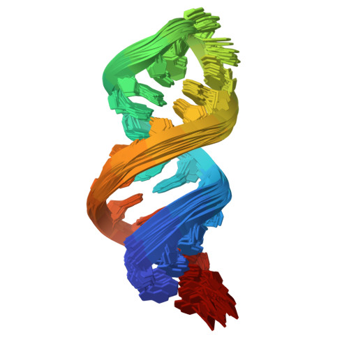
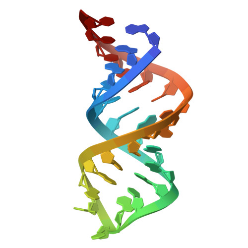
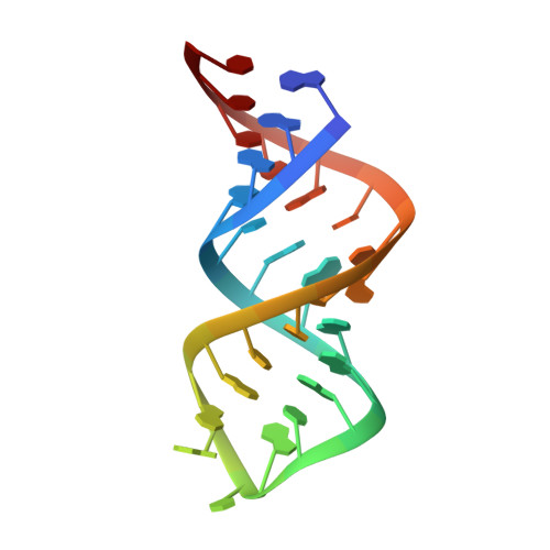
Solution structure of domain 6 from a self-splicing group II intron ribozyme: a Mg(2+) binding site is located close to the stacked branch adenosine.
Erat, M.C., Zerbe, O., Fox, T., Sigel, R.K.O.(2007) Chembiochem 8: 306-314
- PubMed: 17200997
- DOI: https://doi.org/10.1002/cbic.200600459
- Primary Citation of Related Structures:
2AHT - PubMed Abstract:
Group II intron self-splicing is essential for the correct expression of organellar genes in plants, fungi, and yeast, as well as of bacterial genes. Self-excision of these autocatalytic introns from the primary RNA transcript is achieved in a two-step mechanism that is apparently analogous to that of the eukaryotic spliceosome. The 2'-OH of a conserved adenosine (the branch point) located within domain 6 (D6) acts as the nucleophile in the first step of splicing. Despite the biological importance of group II introns, little is known about their structural organization and usage of metal ions in catalysis. Here we report the first solution structure of a catalytically active D6 construct encompassing the branch point and the neighboring helical regions from the mitochondrial yeast intron ai5gamma. The branch adenosine is the single unpaired nucleotide, and, in contrast to the spliceosomal branch site, resides within the helix, being partially stacked between two flanking GU wobble pairs. We identified a novel prominent Mg(2+) binding site in the major groove of the branch site. Importantly, Mg(2+) addition does not impair the stacking of the branch adenosine, rather it strengthens the interaction with the flanking uridines, as shown by NMR and fluorescence studies. This means that domain 6 presents the branch adenosine in a stacked fashion to the core of group II introns upon folding to the active conformation.
Organizational Affiliation:
Institute of Inorganic Chemistry, University of Zürich, Winterthurerstrasse 190, 8057 Zürich, Switzerland.