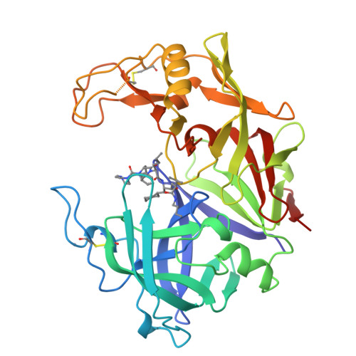
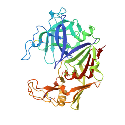
1ZAP

SECRETED ASPARTIC PROTEASE FROM C. ALBICANS


Experimental Data Snapshot

  • Method: X-RAY DIFFRACTION
  • Resolution: 2.50 Å
  • R-Value Work: 0.145 
  • R-Value Observed: 0.145 

wwPDB Validation   3D Report Full Report


This is version 1.4 of the entry. See complete history


Literature

Structure of a secreted aspartic protease from C. albicans complexed with a potent inhibitor: implications for the design of antifungal agents.

Abad-Zapatero, C.Goldman, R.Muchmore, S.W.Hutchins, C.Stewart, K.Navaza, J.Payne, C.D.Ray, T.L.

(1996) Protein Sci 5: 640-652

  • DOI: https://doi.org/10.1002/pro.5560050408
  • Primary Citation of Related Structures:  
    1ZAP

  • PubMed Abstract: 

    The three-dimensional structure of a secreted aspartic protease from Candida albicans complexed with a potent inhibitor reveals variations on the classical aspartic protease theme that dramatically alter the specificity of this class of enzymes. The structure presents: (1) an 8-residue insertion near the first disulfide (Cys 45-Cys 50, pepsin numbering) that results in a broad flap extending toward the active site; (2) a 7-residue deletion replacing helix hN2 (Ser 110-Tyr 114), which enlarges the S3 pocket; (3) a short polar connection between the two rigid body domains that alters their relative orientation and provides certain specificity; and (4) an ordered 11-residue addition at the carboxy terminus. The inhibitor binds in an extended conformation and presents a branched structure at the P3 position. The implications of these findings for the design of potent antifungal agents are discussed.


  • Organizational Affiliation

    Laboratory of Protein Crystallography, Abbott Laboratories, Abbott Park, Illinois 60064-3500, USA.


Macromolecules
Find similar proteins by:  (by identity cutoff)  |  3D Structure
Entity ID: 1
MoleculeChains Sequence LengthOrganismDetailsImage
SECRETED ASPARTIC PROTEINASE342Candida albicansMutation(s): 0 
EC: 3.4.23.24
UniProt
Find proteins for P0CS83 (Candida albicans)
Explore P0CS83 
Go to UniProtKB:  P0CS83
Entity Groups  
Sequence Clusters30% Identity50% Identity70% Identity90% Identity95% Identity100% Identity
UniProt GroupP0CS83
Sequence Annotations
Expand
  • Reference Sequence
Small Molecules
Ligands 2 Unique
IDChains Name / Formula / InChI Key2D Diagram3D Interactions
A70
Query on A70

Download Ideal Coordinates CCD File 
C [auth A]N-ethyl-N-[(4-methylpiperazin-1-yl)carbonyl]-D-phenylalanyl-N-[(1S,2S,4R)-4-(butylcarbamoyl)-1-(cyclohexylmethyl)-2-hyd roxy-5-methylhexyl]-L-norleucinamide
C42 H70 N6 O5
NGCGSAFHWCZMPV-AKHKZFQHSA-N
ZN
Query on ZN

Download Ideal Coordinates CCD File 
B [auth A]ZINC ION
Zn
PTFCDOFLOPIGGS-UHFFFAOYSA-N
Experimental Data & Validation

Experimental Data

  • Method: X-RAY DIFFRACTION
  • Resolution: 2.50 Å
  • R-Value Work: 0.145 
  • R-Value Observed: 0.145 
  • Space Group: P 21 21 21
Unit Cell:
Length ( Å )Angle ( ˚ )
a = 63.6α = 90
b = 98.26β = 90
c = 49.28γ = 90
Software Package:
Software NamePurpose
R-AXISdata collection
DENZOdata reduction
X-PLORmodel building
X-PLORrefinement
R-AXISdata reduction
X-PLORphasing

Structure Validation

View Full Validation Report


Entry History 

Deposition Data

Revision History  (Full details and data files)

  • Version 1.0: 1997-04-21
    Type: Initial release
  • Version 1.1: 2008-03-24
    Changes: Version format compliance
  • Version 1.2: 2011-07-13
    Changes: Non-polymer description, Version format compliance
  • Version 1.3: 2012-01-18
    Changes: Non-polymer description
  • Version 1.4: 2024-10-30
    Changes: Data collection, Database references, Derived calculations, Structure summary