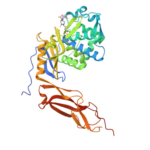
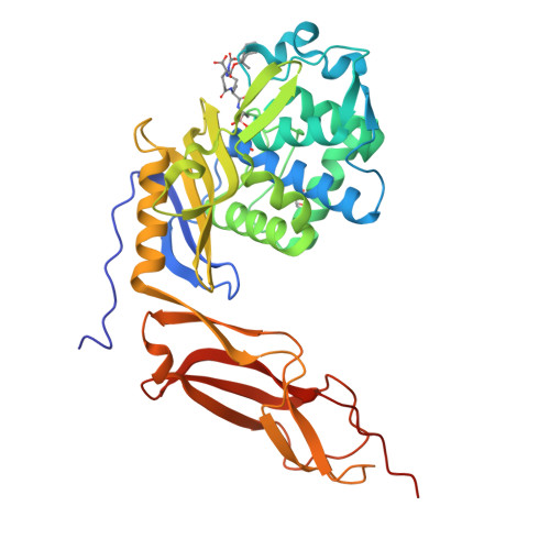
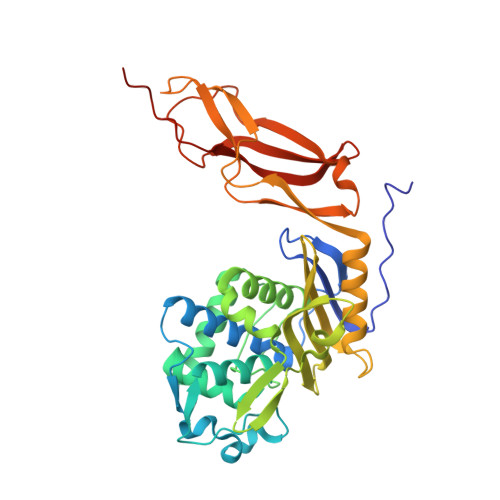
1Z6F

Crystal structure of penicillin-binding protein 5 from E. coli in complex with a boronic acid inhibitor


Experimental Data Snapshot

  • Method: X-RAY DIFFRACTION
  • Resolution: 1.60 Å
  • R-Value Free: 0.240 
  • R-Value Work: 0.213 
  • R-Value Observed: 0.215 

Starting Model: experimental
View more details

wwPDB Validation   3D Report Full Report


Ligand Structure Quality Assessment 


This is version 1.5 of the entry. See complete history


Literature

Crystal Structure of Escherichia coli Penicillin-Binding Protein 5 Bound to a Tripeptide Boronic Acid Inhibitor: A Role for Ser-110 in Deacylation.

Nicola, G.Peddi, S.Stefanova, M.Nicholas, R.A.Gutheil, W.G.Davies, C.

(2005) Biochemistry 44: 8207-8217

  • DOI: https://doi.org/10.1021/bi0473004
  • Primary Citation of Related Structures:  
    1Z6F

  • PubMed Abstract: 

    Penicillin-binding protein 5 (PBP 5) from Escherichia coli is a well-characterized d-alanine carboxypeptidase that serves as a prototypical enzyme to elucidate the structure, function, and catalytic mechanism of PBPs. A comprehensive understanding of the catalytic mechanism underlying d-alanine carboxypeptidation and antibiotic binding has proven elusive. In this study, we report the crystal structure at 1.6 A resolution of PBP 5 in complex with a substrate-like peptide boronic acid, which was designed to resemble the transition-state intermediate during the deacylation step of the enzyme-catalyzed reaction with peptide substrates. In the structure of the complex, the boron atom is covalently attached to Ser-44, which in turn is within hydrogen-bonding distance to Lys-47. This arrangement further supports the assignment of Lys-47 as the general base that activates Ser-44 during acylation. One of the two hydroxyls in the boronyl center (O2) is held by the oxyanion hole comprising the amides of Ser-44 and His-216, while the other hydroxyl (O3), which is analogous to the nucleophilic water for hydrolysis of the acyl-enzyme intermediate, is solvated by a water molecule that bridges to Ser-110. Lys-47 is not well-positioned to act as the catalytic base in the deacylation reaction. Instead, these data suggest a mechanism of catalysis for deacylation that uses a hydrogen-bonding network, involving Lys-213, Ser-110, and a bridging water molecule, to polarize the hydrolytic water molecule.


  • Organizational Affiliation

    Department of Biochemistry, Medical University of South Carolina, Charleston, South Carolina 29425, USA.


Macromolecules
Find similar proteins by:  (by identity cutoff)  |  3D Structure
Entity ID: 1
MoleculeChains Sequence LengthOrganismDetailsImage
Penicillin-binding protein 5363Escherichia coliMutation(s): 0 
Gene Names: dacApfv
EC: 3.4.16.4 (PDB Primary Data), 3.5.2.6 (UniProt)
UniProt
Find proteins for P0AEB2 (Escherichia coli (strain K12))
Explore P0AEB2 
Go to UniProtKB:  P0AEB2
Entity Groups  
Sequence Clusters30% Identity50% Identity70% Identity90% Identity95% Identity100% Identity
UniProt GroupP0AEB2
Sequence Annotations
Expand
  • Reference Sequence
Small Molecules
Ligands 2 Unique
IDChains Name / Formula / InChI Key2D Diagram3D Interactions
BO9
Query on BO9

Download Ideal Coordinates CCD File 
B [auth A]N1-[(1R)-1-(DIHYDROXYBORYL)ETHYL]-N2-[(TERT-BUTOXYCARBONYL)-D-GAMMA-GLUTAMYL]-N6-[(BENZYLOXY)CARBONYL-L-LYSINAMIDE
C26 H41 B N4 O10
OTTYWVUOPWENAJ-HOJAQTOUSA-N
GOL
Query on GOL

Download Ideal Coordinates CCD File 
C [auth A]GLYCEROL
C3 H8 O3
PEDCQBHIVMGVHV-UHFFFAOYSA-N
Binding Affinity Annotations 
IDSourceBinding Affinity
BO9 PDBBind:  1Z6F Ki: 1.30e+4 (nM) from 1 assay(s)
Experimental Data & Validation

Experimental Data

  • Method: X-RAY DIFFRACTION
  • Resolution: 1.60 Å
  • R-Value Free: 0.240 
  • R-Value Work: 0.213 
  • R-Value Observed: 0.215 
  • Space Group: C 1 2 1
Unit Cell:
Length ( Å )Angle ( ˚ )
a = 109.76α = 90
b = 50.25β = 120.2
c = 84.31γ = 90
Software Package:
Software NamePurpose
REFMACrefinement
MAR345data collection
SCALEPACKdata scaling

Structure Validation

View Full Validation Report


Ligand Structure Quality Assessment 


Entry History 

Deposition Data

Revision History  (Full details and data files)

  • Version 1.0: 2005-06-21
    Type: Initial release
  • Version 1.1: 2008-04-30
    Changes: Version format compliance
  • Version 1.2: 2011-07-13
    Changes: Non-polymer description, Version format compliance
  • Version 1.3: 2017-10-11
    Changes: Advisory, Refinement description
  • Version 1.4: 2023-08-23
    Changes: Advisory, Data collection, Database references, Derived calculations, Refinement description
  • Version 1.5: 2024-11-20
    Changes: Structure summary