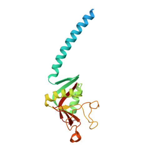
1PWB

High resolution crystal structure of an active recombinant fragment of human lung surfactant protein D with maltose


Experimental Data Snapshot

  • Method: X-RAY DIFFRACTION
  • Resolution: 1.40 Å
  • R-Value Free: 0.228 
  • R-Value Work: 0.205 
  • R-Value Observed: 0.205 

Starting Model: experimental
View more details

wwPDB Validation   3D Report Full Report


Ligand Structure Quality Assessment 


This is version 2.3 of the entry. See complete history


Literature

High-resolution structural insights into ligand binding and immune cell recognition by human lung surfactant protein D

Shrive, A.K.Tharia, H.A.Strong, P.Kishore, U.Burns, I.Rizkallah, P.J.Reid, K.B.Greenhough, T.J.

(2003) J Mol Biol 331: 509-523

  • DOI: https://doi.org/10.1016/s0022-2836(03)00761-7
  • Primary Citation of Related Structures:  
    1PW9, 1PWB

  • PubMed Abstract: 

    Lung surfactant protein D (SP-D) can directly interact with carbohydrate residues on pulmonary pathogens and allergens, stimulate immune cells, and manipulate cytokine and chemokine profiles during the immune response in the lungs. Therapeutic administration of rfhSP-D, a recombinant homotrimeric fragment of human SP-D comprising the alpha-helical coiled-coil neck plus three CRDs, protects mice against lung allergy and infection caused by the fungal pathogen Aspergillus fumigatus. The high resolution crystal structures of maltose-bound rfhSP-D to 1.4A, and of rfhSP-D to 1.6A, define the fine detail of the mode and nature of carbohydrate recognition and provide insights into how a small fragment of human SP-D can bind to allergens/antigens or whole pathogens, and at the same time recruit and engage effector cells and molecules of humoral immunity. A previously unreported calcium ion, located on the trimeric axis in a pore at the bottom of the funnel formed by the three CRDs and close to the neck-CRD interface, is coordinated by a triad of glutamate residues which are, to some extent, neutralised by their interactions with a triad of exposed lysine residues in the funnel. The spatial relationship between the neck and the CRDs is maintained internally by these lysine residues, and externally by a glutamine, which forms a pair of hydrogen-bonds within an external cleft at each neck-CRD interface. Structural links between the central pore and the cleft suggest a possible effector mechanism for immune cell surface receptor binding in the presence of bound, extended natural lipopolysaccharide and phospholipid ligands. The structural requirements for such an effector mechanism, involving both the trimeric framework for multivalent ligand binding and recognition sites formed from more than one subunit, are present in both native hSP-D and rfhSP-D, providing a possible explanation for the significant biological activity of rfhSP-D.


  • Organizational Affiliation

    School of Life Sciences, Keele University, Staffordshire ST5 5BG, Keele, UK. [email protected]


Macromolecules
Find similar proteins by:  (by identity cutoff)  |  3D Structure
Entity ID: 1
MoleculeChains Sequence LengthOrganismDetailsImage
Pulmonary surfactant-associated protein D
A, B, C
177Homo sapiensMutation(s): 1 
UniProt & NIH Common Fund Data Resources
Find proteins for P35247 (Homo sapiens)
Explore P35247 
Go to UniProtKB:  P35247
PHAROS:  P35247
GTEx:  ENSG00000133661 
Entity Groups  
Sequence Clusters30% Identity50% Identity70% Identity90% Identity95% Identity100% Identity
UniProt GroupP35247
Sequence Annotations
Expand
  • Reference Sequence
Oligosaccharides

Help

Entity ID: 2
MoleculeChains Length2D Diagram Glycosylation3D Interactions
alpha-D-glucopyranose-(1-4)-alpha-D-glucopyranose
D
2N/A
Glycosylation Resources
GlyTouCan:  G07411ON
GlyCosmos:  G07411ON
Small Molecules
Ligands 2 Unique
IDChains Name / Formula / InChI Key2D Diagram3D Interactions
GLC
Query on GLC

Download Ideal Coordinates CCD File 
H [auth B],
L [auth C]
alpha-D-glucopyranose
C6 H12 O6
WQZGKKKJIJFFOK-DVKNGEFBSA-N
CA
Query on CA

Download Ideal Coordinates CCD File 
E [auth A]
F [auth A]
G [auth A]
I [auth B]
J [auth B]
E [auth A],
F [auth A],
G [auth A],
I [auth B],
J [auth B],
K [auth B],
M [auth C],
N [auth C],
O [auth C]
CALCIUM ION
Ca
BHPQYMZQTOCNFJ-UHFFFAOYSA-N
Biologically Interesting Molecules (External Reference) 1 Unique
Experimental Data & Validation

Experimental Data

  • Method: X-RAY DIFFRACTION
  • Resolution: 1.40 Å
  • R-Value Free: 0.228 
  • R-Value Work: 0.205 
  • R-Value Observed: 0.205 
  • Space Group: P 1 21 1
Unit Cell:
Length ( Å )Angle ( ˚ )
a = 55.48α = 90
b = 108.3β = 91.43
c = 55.78γ = 90
Software Package:
Software NamePurpose
MOSFLMdata reduction
SCALAdata scaling
X-PLORmodel building
X-PLORrefinement
CCP4data scaling
X-PLORphasing

Structure Validation

View Full Validation Report


Ligand Structure Quality Assessment 


Entry History 

Deposition Data

Revision History  (Full details and data files)

  • Version 1.0: 2003-08-05
    Type: Initial release
  • Version 1.1: 2008-04-29
    Changes: Version format compliance
  • Version 1.2: 2011-07-13
    Changes: Version format compliance
  • Version 2.0: 2020-07-29
    Type: Remediation
    Reason: Carbohydrate remediation
    Changes: Atomic model, Data collection, Derived calculations, Structure summary
  • Version 2.1: 2021-10-27
    Changes: Database references, Structure summary
  • Version 2.2: 2023-08-16
    Changes: Data collection, Refinement description
  • Version 2.3: 2024-11-13
    Changes: Structure summary