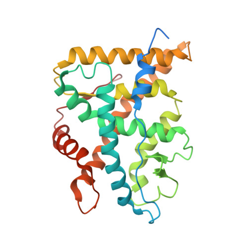
1P93

CRYSTAL STRUCTURE OF THE AGONIST FORM OF GLUCOCORTICOID RECEPTOR


Experimental Data Snapshot

  • Method: X-RAY DIFFRACTION
  • Resolution: 2.70 Å
  • R-Value Free: 0.363 
  • R-Value Work: 0.345 
  • R-Value Observed: 0.350 

Starting Model: experimental
View more details

wwPDB Validation   3D Report Full Report


This is version 1.6 of the entry. See complete history


Literature

The Three-dimensional Structures of Antagonistic and Agonistic Forms of the Glucocorticoid Receptor Ligand-binding Domain: RU-486 INDUCES A TRANSCONFORMATION THAT LEADS TO ACTIVE ANTAGONISM.

Kauppi, B.Jakob, C.Farnegardh, M.Yang, J.Ahola, H.Alarcon, M.Calles, K.Engstrom, O.Harlan, J.Muchmore, S.Ramqvist, A.-K.Thorell, S.Ohman, L.Greer, J.Gustafsson, J.-A.Carlstedt-Duke, J.Carlquist, M.

(2003) J Biol Chem 278: 22748-22754

  • DOI: https://doi.org/10.1074/jbc.M212711200
  • Primary Citation of Related Structures:  
    1NHZ, 1P93

  • PubMed Abstract: 

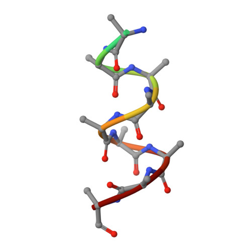
    Here we describe the three-dimensional crystal structures of human glucocorticoid receptor ligand-binding domain (GR-LBD) in complex with the antagonist RU-486 at 2.3 A resolution and with the agonist dexamethasone ligand together with a coactivator peptide at 2.8 A. The RU-486 structure was solved in several different crystal forms, two with helix 12 intact (GR1 and GR3) and one with a protease-digested C terminus (GR2). In GR1, part of helix 12 is in a position that covers the co-activator pocket, whereas in the GR3, domain swapping is seen between the crystallographically identical subunits in the GR dimer. An arm consisting of the end of helix 11 and beyond stretches out from one molecule, and helix 12 binds to the other LBD, partly blocking the coactivator pocket of that molecule. This type of GR-LBD dimer has not been described before but might be an artifact from crystallization. Furthermore, the subunits of the GR3 dimers are covalently connected via a disulfide bond between the Cys-736 residues in the two molecules. All three RU-486 GR-LBD structures show that GR has a very flexible region between the end of helix 11 and the end of helix 12.


  • Organizational Affiliation

    Structure Biology, Karo Bio AB, Novum, SE-141 57 Huddinge, Sweden. [email protected]


Macromolecules
Find similar proteins by:  (by identity cutoff)  |  3D Structure
Entity ID: 1
MoleculeChains Sequence LengthOrganismDetailsImage
Glucocorticoid receptorA,
C [auth B],
E [auth C],
G [auth D]
280Homo sapiensMutation(s): 3 
Gene Names: NR3C1 or GRL
UniProt & NIH Common Fund Data Resources
Find proteins for P04150 (Homo sapiens)
Explore P04150 
Go to UniProtKB:  P04150
PHAROS:  P04150
GTEx:  ENSG00000113580 
Entity Groups  
Sequence Clusters30% Identity50% Identity70% Identity90% Identity95% Identity100% Identity
UniProt GroupP04150
Sequence Annotations
Expand
  • Reference Sequence

Find similar proteins by:  Sequence   |   3D Structure  

Entity ID: 2
MoleculeChains Sequence LengthOrganismDetailsImage
Nuclear receptor coactivator 2B [auth E],
D [auth F],
F [auth G],
H
12N/AMutation(s): 0 
UniProt & NIH Common Fund Data Resources
Find proteins for Q15596 (Homo sapiens)
Explore Q15596 
Go to UniProtKB:  Q15596
PHAROS:  Q15596
GTEx:  ENSG00000140396 
Entity Groups  
Sequence Clusters30% Identity50% Identity70% Identity90% Identity95% Identity100% Identity
UniProt GroupQ15596
Sequence Annotations
Expand
  • Reference Sequence
Small Molecules
Binding Affinity Annotations 
IDSourceBinding Affinity
DEX BindingDB:  1P93 Ki: min: 0.69, max: 2900 (nM) from 10 assay(s)
Kd: min: 2, max: 19 (nM) from 3 assay(s)
IC50: min: 0.5, max: 22 (nM) from 33 assay(s)
EC50: min: 0.1, max: 30 (nM) from 22 assay(s)
PDBBind:  1P93 Kd: 19 (nM) from 1 assay(s)
Experimental Data & Validation

Experimental Data

  • Method: X-RAY DIFFRACTION
  • Resolution: 2.70 Å
  • R-Value Free: 0.363 
  • R-Value Work: 0.345 
  • R-Value Observed: 0.350 
  • Space Group: P 31
Unit Cell:
Length ( Å )Angle ( ˚ )
a = 127.4α = 90
b = 127.4β = 90
c = 91.8γ = 120
Software Package:
Software NamePurpose
CNXrefinement
MOSFLMdata reduction
CCP4data scaling
MOLREPphasing

Structure Validation

View Full Validation Report


Entry History 

Revision History  (Full details and data files)

  • Version 1.0: 2003-07-08
    Type: Initial release
  • Version 1.1: 2008-04-29
    Changes: Version format compliance
  • Version 1.2: 2011-07-13
    Changes: Version format compliance
  • Version 1.3: 2018-01-31
    Changes: Experimental preparation
  • Version 1.4: 2020-09-02
    Changes: Data collection, Database references, Derived calculations, Structure summary
  • Version 1.5: 2021-10-27
    Changes: Database references
  • Version 1.6: 2023-08-16
    Changes: Data collection, Refinement description