1O03

Structure of Pentavalent Phosphorous Intermediate of an Enzyme Catalyzed Phosphoryl transfer Reaction observed on cocrystallization with Glucose 6-phosphate


Experimental Data Snapshot

  • Method: X-RAY DIFFRACTION
  • Resolution: 1.40 Å
  • R-Value Free: 0.194 
  • R-Value Work: 0.170 
  • R-Value Observed: 0.220 

Starting Model: experimental
View more details

wwPDB Validation   3D Report Full Report


Ligand Structure Quality Assessment 

Created with Raphaël 2.3.0Worse 01 BetterLigand structure goodness of fit to experimental dataBest fitted G16Click on this verticalbar to view details

This is version 1.5 of the entry. See complete history


Literature

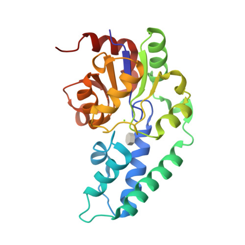
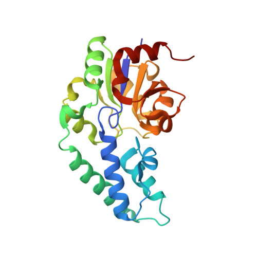
The pentacovalent phosphorus intermediate of a phosphoryl transfer reaction.

Lahiri, S.D.Zhang, G.Dunaway-Mariano, D.Allen, K.N.

(2003) Science 299: 2067-2071

  • DOI: https://doi.org/10.1126/science.1082710
  • Primary Citation of Related Structures:  
    1O03, 1O08

  • PubMed Abstract: 

    Enzymes provide enormous rate enhancements, unmatched by any other type of catalyst. The stabilization of high-energy states along the reaction coordinate is the crux of the catalytic power of enzymes. We report the atomic-resolution structure of a high-energy reaction intermediate stabilized in the active site of an enzyme. Crystallization of phosphorylated beta-phosphoglucomutase in the presence of the Mg(II) cofactor and either of the substrates glucose 1-phosphate or glucose 6-phosphate produced crystals of the enzyme-Mg(II)-glucose 1,6-(bis)phosphate complex, which diffracted x-rays to 1.2 and 1.4 angstroms, respectively. The structure reveals a stabilized pentacovalent phosphorane formed in the phosphoryl transfer from the C(1)O of glucose 1,6-(bis)phosphate to the nucleophilic Asp8 carboxylate.


  • Organizational Affiliation

    Department of Physiology and Biophysics, Boston University School of Medicine, Boston, MA 02118-2394, USA.


Macromolecules
Find similar proteins by:  (by identity cutoff)  |  3D Structure
Entity ID: 1
MoleculeChains Sequence LengthOrganismDetailsImage
beta-phosphoglucomutase221Lactococcus lactisMutation(s): 0 
EC: 5.4.2.6
UniProt
Find proteins for P71447 (Lactococcus lactis subsp. lactis (strain IL1403))
Explore P71447 
Go to UniProtKB:  P71447
Entity Groups  
Sequence Clusters30% Identity50% Identity70% Identity90% Identity95% Identity100% Identity
UniProt GroupP71447
Sequence Annotations
Expand
  • Reference Sequence
Experimental Data & Validation

Experimental Data

  • Method: X-RAY DIFFRACTION
  • Resolution: 1.40 Å
  • R-Value Free: 0.194 
  • R-Value Work: 0.170 
  • R-Value Observed: 0.220 
  • Space Group: P 21 21 21
Unit Cell:
Length ( Å )Angle ( ˚ )
a = 37.311α = 90
b = 54.173β = 90
c = 104.446γ = 90
Software Package:
Software NamePurpose
DENZOdata reduction
SCALEPACKdata scaling
AMoREphasing
CNSrefinement

Structure Validation

View Full Validation Report


Ligand Structure Quality Assessment 

Created with Raphaël 2.3.0Worse 01 BetterLigand structure goodness of fit to experimental dataBest fitted G16Click on this verticalbar to view details

Entry History 

Deposition Data

Revision History  (Full details and data files)

  • Version 1.0: 2003-03-18
    Type: Initial release
  • Version 1.1: 2008-04-29
    Changes: Version format compliance
  • Version 1.2: 2011-07-13
    Changes: Version format compliance
  • Version 1.3: 2020-07-29
    Type: Remediation
    Reason: Carbohydrate remediation
    Changes: Advisory, Data collection, Derived calculations, Structure summary
  • Version 1.4: 2024-02-14
    Changes: Data collection, Database references, Structure summary
  • Version 1.5: 2024-04-03
    Changes: Refinement description