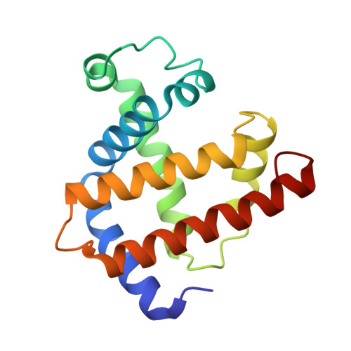
1MBC

X-RAY STRUCTURE AND REFINEMENT OF CARBON-MONOXY (FE II)-MYOGLOBIN AT 1.5 ANGSTROMS RESOLUTION


Experimental Data Snapshot

  • Method: X-RAY DIFFRACTION
  • Resolution: 1.50 Å
  • R-Value Observed: 0.171 

wwPDB Validation   3D Report Full Report


This is version 1.3 of the entry. See complete history


Literature

X-ray structure and refinement of carbon-monoxy (Fe II)-myoglobin at 1.5 A resolution.

Kuriyan, J.Wilz, S.Karplus, M.Petsko, G.A.

(1986) J Mol Biol 192: 133-154

  • DOI: https://doi.org/10.1016/0022-2836(86)90470-5
  • Primary Citation of Related Structures:  
    1MBC

  • PubMed Abstract: 

    The structure of carbon-monoxy (Fe II) myoglobin at 260 K has been solved at a resolution of 1.5 A by X-ray diffraction and a model refined against the X-ray data by restrained least-squares. The CO ligand is disordered and distorted from the linear conformation seen in model compounds. At least two conformations, with Fe--C--O angles of 140 degrees and 120 degrees, are required to model the system. The heme pocket is significantly larger than in deoxy-myoglobin because the distal residues have relaxed around the ligand; the largest displacement occurs for the distal histidine side-chain, which moves more than 1.4 A on ligand binding. The side-chain of Arg45 (CD3) is disordered and apparently exists in two equally populated conformations. One of these does not block the motion of the distal histidine out of the binding pocket, suggesting a mechanism for ligand entry. The heme group is planar (root-mean-square deviation from planarity is 0.08 A) with no doming of the pyrrole groups. The Fe--N epsilon 2 (His93) bond length is 2.2 A and the Fe--C bond length in the CO complex is 1.9 A. The iron is the least-squares plane of the heme, and this leads to the proximal histidine moving by 0.4 A relative to its position in deoxy-myoglobin. This shift correlates with a global structural change, with the proximal part of the molecule translated towards the heme plane.


Macromolecules
Find similar proteins by:  (by identity cutoff)  |  3D Structure
Entity ID: 1
MoleculeChains Sequence LengthOrganismDetailsImage
MYOGLOBIN153Physeter macrocephalusMutation(s): 0 
EC: 1.11.1 (UniProt), 1.7 (UniProt)
UniProt
Find proteins for P02185 (Physeter macrocephalus)
Explore P02185 
Go to UniProtKB:  P02185
Entity Groups  
Sequence Clusters30% Identity50% Identity70% Identity90% Identity95% Identity100% Identity
UniProt GroupP02185
Sequence Annotations
Expand
  • Reference Sequence
Small Molecules
Ligands 3 Unique
IDChains Name / Formula / InChI Key2D Diagram3D Interactions
HEM
Query on HEM

Download Ideal Coordinates CCD File 
C [auth A]PROTOPORPHYRIN IX CONTAINING FE
C34 H32 Fe N4 O4
KABFMIBPWCXCRK-RGGAHWMASA-L
SO4
Query on SO4

Download Ideal Coordinates CCD File 
B [auth A]SULFATE ION
O4 S
QAOWNCQODCNURD-UHFFFAOYSA-L
CMO
Query on CMO

Download Ideal Coordinates CCD File 
D [auth A]CARBON MONOXIDE
C O
UGFAIRIUMAVXCW-UHFFFAOYSA-N
Experimental Data & Validation

Experimental Data

  • Method: X-RAY DIFFRACTION
  • Resolution: 1.50 Å
  • R-Value Observed: 0.171 
  • Space Group: P 1 21 1
Unit Cell:
Length ( Å )Angle ( ˚ )
a = 64.18α = 90
b = 30.84β = 105.84
c = 34.69γ = 90
Software Package:
Software NamePurpose
PROLSQrefinement

Structure Validation

View Full Validation Report


Entry History 

Deposition Data

Revision History  (Full details and data files)

  • Version 1.0: 1989-01-09
    Type: Initial release
  • Version 1.1: 2008-03-24
    Changes: Version format compliance
  • Version 1.2: 2011-07-13
    Changes: Version format compliance
  • Version 1.3: 2024-02-14
    Changes: Data collection, Database references, Derived calculations, Other