1J7D

Crystal Structure of hMms2-hUbc13


Experimental Data Snapshot

  • Method: X-RAY DIFFRACTION
  • Resolution: 1.85 Å
  • R-Value Free: 
    0.244 (Depositor), 0.210 (DCC) 
  • R-Value Work: 
    0.215 (Depositor), 0.210 (DCC) 

wwPDB Validation   3D Report Full Report


This is version 1.3 of the entry. See complete history


Literature

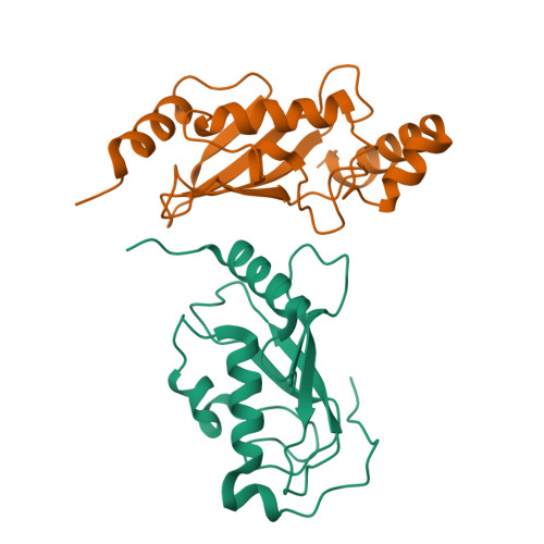
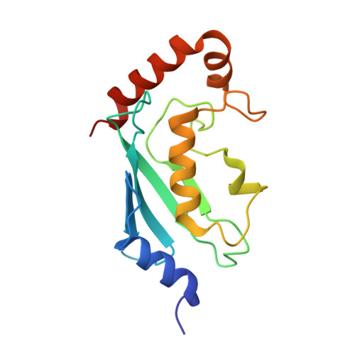
Crystal structure of the human ubiquitin conjugating enzyme complex, hMms2-hUbc13.

Moraes, T.F.Edwards, R.A.McKenna, S.Pastushok, L.Xiao, W.Glover, J.N.Ellison, M.J.

(2001) Nat Struct Biol 8: 669-673

  • DOI: https://doi.org/10.1038/90373
  • Primary Citation of Related Structures:  
    1J74, 1J7D

  • PubMed Abstract: 

    The ubiquitin conjugating enzyme complex Mms2-Ubc13 plays a key role in post-replicative DNA repair in yeast and the NF-kappaB signal transduction pathway in humans. This complex assembles novel polyubiquitin chains onto yet uncharacterized protein targets. Here we report the crystal structure of a complex between hMms2 (Uev1) and hUbc13 at 1.85 A resolution and a structure of free hMms2 at 1.9 A resolution. These structures reveal that the hMms2 monomer undergoes a localized conformational change upon interaction with hUbc13. The nature of the interface provides a physical basis for the preference of Mms2 for Ubc13 as a partner over a variety of other structurally similar ubiquitin-conjugating enzymes. The structure of the hMms2-hUbc13 complex provides the conceptual foundation for understanding the mechanism of Lys 63 multiubiquitin chain assembly and for its interactions with the RING finger proteins Rad5 and Traf6.


  • Organizational Affiliation

    Department of Biochemistry, University of Alberta, Edmonton, Alberta, T6G-2H7, Canada.


Macromolecules
Find similar proteins by:  (by identity cutoff)  |  3D Structure
Entity ID: 1
MoleculeChains Sequence LengthOrganismDetailsImage
MMS2145Homo sapiensMutation(s): 0 
UniProt & NIH Common Fund Data Resources
Find proteins for Q15819 (Homo sapiens)
Explore Q15819 
Go to UniProtKB:  Q15819
PHAROS:  Q15819
GTEx:  ENSG00000169139 
Entity Groups  
Sequence Clusters30% Identity50% Identity70% Identity90% Identity95% Identity100% Identity
UniProt GroupQ15819
Sequence Annotations
Expand
  • Reference Sequence
Find similar proteins by:  (by identity cutoff)  |  3D Structure
Entity ID: 2
MoleculeChains Sequence LengthOrganismDetailsImage
UBIQUITIN-CONJUGATING ENZYME E2-17 KDA152Homo sapiensMutation(s): 0 
EC: 6.3.2.19 (PDB Primary Data), 2.3.2.23 (UniProt)
UniProt & NIH Common Fund Data Resources
Find proteins for P61088 (Homo sapiens)
Explore P61088 
Go to UniProtKB:  P61088
PHAROS:  P61088
GTEx:  ENSG00000177889 
Entity Groups  
Sequence Clusters30% Identity50% Identity70% Identity90% Identity95% Identity100% Identity
UniProt GroupP61088
Sequence Annotations
Expand
  • Reference Sequence
Experimental Data & Validation

Experimental Data

  • Method: X-RAY DIFFRACTION
  • Resolution: 1.85 Å
  • R-Value Free:  0.244 (Depositor), 0.210 (DCC) 
  • R-Value Work:  0.215 (Depositor), 0.210 (DCC) 
Space Group: P 21 21 21
Unit Cell:
Length ( Å )Angle ( ˚ )
a = 44.132α = 90
b = 74.137β = 90
c = 91.489γ = 90
Software Package:
Software NamePurpose
DENZOdata reduction
SCALEPACKdata scaling
SOLVEphasing
CNSrefinement

Structure Validation

View Full Validation Report


Entry History 

Deposition Data

Revision History  (Full details and data files)

  • Version 1.0: 2001-08-08
    Type: Initial release
  • Version 1.1: 2008-04-27
    Changes: Version format compliance
  • Version 1.2: 2011-07-13
    Changes: Version format compliance
  • Version 1.3: 2024-02-07
    Changes: Data collection, Database references