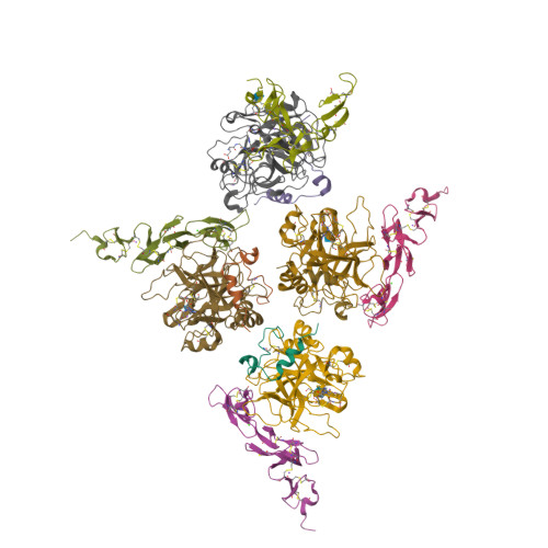
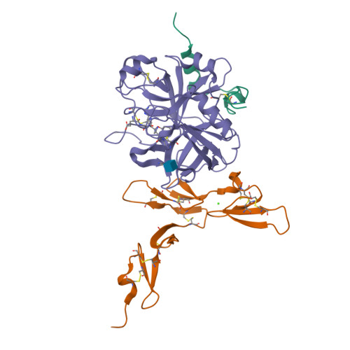
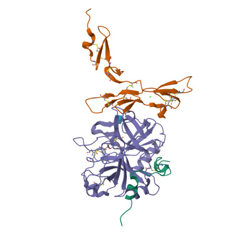
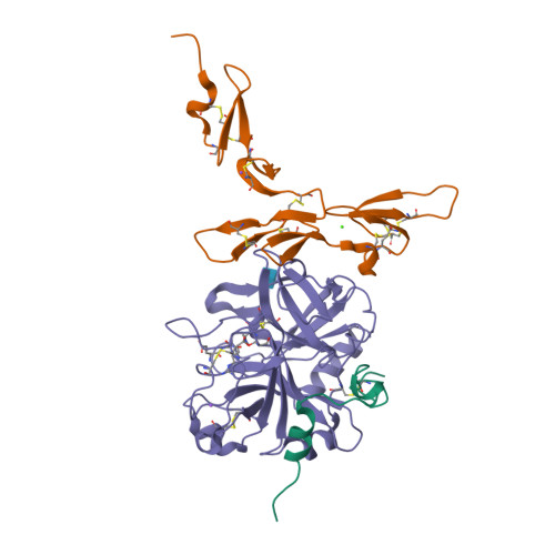
1DX5

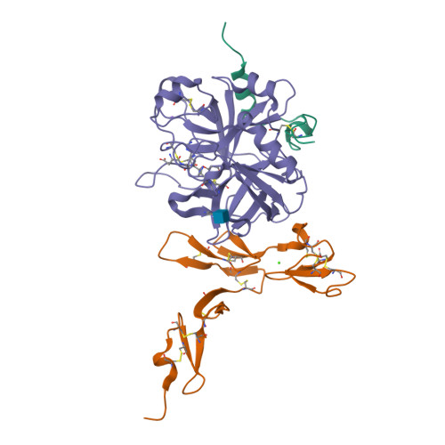
Crystal structure of the thrombin-thrombomodulin complex


Experimental Data Snapshot

  • Method: X-RAY DIFFRACTION
  • Resolution: 2.30 Å
  • R-Value Free: 0.241 
  • R-Value Work: 0.200 
  • R-Value Observed: 0.200 

Starting Model: other
View more details

wwPDB Validation   3D Report Full Report


This is version 2.3 of the entry. See complete history


Literature

Structural Basis for the Anticoagulant Activity of the Thrombin-Thrombomodulin Complex

Fuentes-Prior, P.Iwanaga, Y.Huber, R.Pagila, R.Rumennik, G.Seto, M.Morser, J.Light, D.R.Bode, W.

(2000) Nature 404: 518

  • DOI: https://doi.org/10.1038/35006683
  • Primary Citation of Related Structures:  
    1DX5

  • PubMed Abstract: 

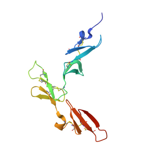
    The serine proteinase alpha-thrombin causes blood clotting through proteolytic cleavage of fibrinogen and protease-activated receptors and amplifies its own generation by activating the essential clotting factors V and VIII. Thrombomodulin, a transmembrane thrombin receptor with six contiguous epidermal growth factor-like domains (TME1-6), profoundly alters the substrate specificity of thrombin from pro- to anticoagulant by activating protein C. Activated protein C then deactivates the coagulation cascade by degrading activated factors V and VIII. The thrombin-thrombomodulin complex inhibits fibrinolysis by activating the procarboxypeptidase thrombin-activatable fibrinolysis inhibitor. Here we present the 2.3 A crystal structure of human alpha-thrombin bound to the smallest thrombomodulin fragment required for full protein-C co-factor activity, TME456. The Y-shaped thrombomodulin fragment binds to thrombin's anion-binding exosite-I, preventing binding of procoagulant substrates. Thrombomodulin binding does not seem to induce marked allosteric structural rearrangements at the thrombin active site. Rather, docking of a protein C model to thrombin-TME456 indicates that TME45 may bind substrates in such a manner that their zymogen-activation cleavage sites are presented optimally to the unaltered thrombin active site.


  • Organizational Affiliation

    Max-Planck-Institut für Biochemie, Martinsried, Germany. [email protected]


Macromolecules
Find similar proteins by:  (by identity cutoff)  |  3D Structure
Entity ID: 1
MoleculeChains Sequence LengthOrganismDetailsImage
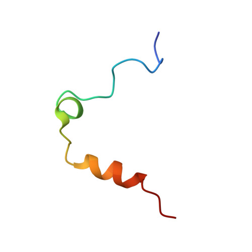
Thrombin light chain
A, B, C, D
36Homo sapiensMutation(s): 0 
EC: 3.4.21.5
UniProt & NIH Common Fund Data Resources
Find proteins for P00734 (Homo sapiens)
Explore P00734 
Go to UniProtKB:  P00734
PHAROS:  P00734
GTEx:  ENSG00000180210 
Entity Groups  
Sequence Clusters30% Identity50% Identity70% Identity90% Identity95% Identity100% Identity
UniProt GroupP00734
Sequence Annotations
Expand
  • Reference Sequence
Find similar proteins by:  (by identity cutoff)  |  3D Structure
Entity ID: 2
MoleculeChains Sequence LengthOrganismDetailsImage
ThrombomodulinE [auth I],
F [auth J],
G [auth K],
H [auth L]
118Homo sapiensMutation(s): 0 
Gene Names: THBDTHRM
UniProt & NIH Common Fund Data Resources
Find proteins for P07204 (Homo sapiens)
Explore P07204 
Go to UniProtKB:  P07204
PHAROS:  P07204
GTEx:  ENSG00000178726 
Entity Groups  
Sequence Clusters30% Identity50% Identity70% Identity90% Identity95% Identity100% Identity
UniProt GroupP07204
Sequence Annotations
Expand
  • Reference Sequence
Find similar proteins by:  (by identity cutoff)  |  3D Structure
Entity ID: 3
MoleculeChains Sequence LengthOrganismDetailsImage
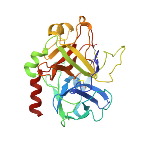
Thrombin heavy chainI [auth M],
J [auth N],
K [auth O],
L [auth P]
259Homo sapiensMutation(s): 0 
EC: 3.4.21.5
UniProt & NIH Common Fund Data Resources
Find proteins for P00734 (Homo sapiens)
Explore P00734 
Go to UniProtKB:  P00734
PHAROS:  P00734
GTEx:  ENSG00000180210 
Entity Groups  
Sequence Clusters30% Identity50% Identity70% Identity90% Identity95% Identity100% Identity
UniProt GroupP00734
Glycosylation
Glycosylation Sites: 1Go to GlyGen: P00734-1
Sequence Annotations
Expand
  • Reference Sequence
Small Molecules
Ligands 5 Unique
IDChains Name / Formula / InChI Key2D Diagram3D Interactions
0GJ
Query on 0GJ

Download Ideal Coordinates CCD File 
AA [auth M]
BA [auth M]
CA [auth M]
DA [auth M]
GA [auth N]
AA [auth M],
BA [auth M],
CA [auth M],
DA [auth M],
GA [auth N],
HA [auth N],
IA [auth N],
JA [auth N],
MA [auth O],
NA [auth O],
OA [auth O],
PA [auth O],
TA [auth P],
UA [auth P],
VA [auth P],
WA [auth P]
L-alpha-glutamyl-N-{(1S)-4-{[amino(iminio)methyl]amino}-1-[(1S)-2-chloro-1-hydroxyethyl]butyl}glycinamide
C14 H28 Cl N6 O5
XELWNHKFCNMWQO-LPEHRKFASA-O
NAG
Query on NAG

Download Ideal Coordinates CCD File 
EA [auth N],
KA [auth O],
QA [auth P],
Y [auth M]
2-acetamido-2-deoxy-beta-D-glucopyranose
C8 H15 N O6
OVRNDRQMDRJTHS-FMDGEEDCSA-N
FMT
Query on FMT

Download Ideal Coordinates CCD File 
M [auth I],
P [auth J],
S [auth K],
SA [auth P],
V [auth L]
FORMIC ACID
C H2 O2
BDAGIHXWWSANSR-UHFFFAOYSA-N
CA
Query on CA

Download Ideal Coordinates CCD File 
N [auth I],
Q [auth J],
T [auth K],
W [auth L]
CALCIUM ION
Ca
BHPQYMZQTOCNFJ-UHFFFAOYSA-N
NA
Query on NA

Download Ideal Coordinates CCD File 
FA [auth N]
LA [auth O]
O [auth I]
R [auth J]
RA [auth P]
FA [auth N],
LA [auth O],
O [auth I],
R [auth J],
RA [auth P],
U [auth K],
X [auth L],
Z [auth M]
SODIUM ION
Na
FKNQFGJONOIPTF-UHFFFAOYSA-N
Biologically Interesting Molecules (External Reference) 1 Unique
Entity ID: 8
IDChains NameType/Class2D Diagram3D Interactions
PRD_000288 (0GJ)
Query on PRD_000288
AA [auth M]
BA [auth M]
CA [auth M]
DA [auth M]
GA [auth N]
AA [auth M], BA [auth M], CA [auth M], DA [auth M], GA [auth N], HA [auth N], IA [auth N], JA [auth N], MA [auth O], NA [auth O], OA [auth O], PA [auth O], TA [auth P], UA [auth P], VA [auth P], WA [auth P]
GLU-GLY-ARG-CHLOROMETHYL KETONEPeptide-like / Inhibitor
Experimental Data & Validation

Experimental Data

  • Method: X-RAY DIFFRACTION
  • Resolution: 2.30 Å
  • R-Value Free: 0.241 
  • R-Value Work: 0.200 
  • R-Value Observed: 0.200 
  • Space Group: H 3
Unit Cell:
Length ( Å )Angle ( ˚ )
a = 214.4α = 90
b = 214.4β = 90
c = 131.41γ = 120
Software Package:
Software NamePurpose
X-PLORrefinement
MOSFLMdata reduction
SCALAdata scaling
X-PLORphasing

Structure Validation

View Full Validation Report


Entry History 

Deposition Data

Revision History  (Full details and data files)

  • Version 1.0: 2000-04-10
    Type: Initial release
  • Version 1.1: 2011-07-13
    Changes: Atomic model, Database references, Derived calculations, Non-polymer description, Structure summary, Version format compliance
  • Version 1.2: 2016-12-21
    Changes: Advisory, Atomic model, Derived calculations, Non-polymer description, Other, Source and taxonomy
  • Version 2.0: 2018-06-27
    Changes: Advisory, Atomic model, Data collection, Database references, Derived calculations, Non-polymer description, Polymer sequence, Source and taxonomy, Structure summary
  • Version 2.1: 2020-07-29
    Type: Remediation
    Reason: Carbohydrate remediation
    Changes: Data collection, Derived calculations, Structure summary
  • Version 2.2: 2024-05-01
    Changes: Advisory, Data collection, Database references, Refinement description, Structure summary
  • Version 2.3: 2024-11-13
    Changes: Structure summary