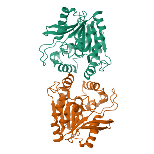
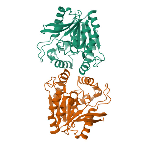
Crystal structure of the secreted form of antigen 85C reveals potential targets for mycobacterial drugs and vaccines.
Ronning, D.R., Klabunde, T., Besra, G.S., Vissa, V.D., Belisle, J.T., Sacchettini, J.C.(2000) Nat Struct Biol 7: 141-146
- PubMed: 10655617
- DOI: https://doi.org/10.1038/72413
- Primary Citation of Related Structures:
1DQY, 1DQZ - PubMed Abstract:
The antigen 85 (ag85) complex, composed of three proteins (ag85A, B and C), is a major protein component of the Mycobacterium tuberculosis cell wall. Each protein possesses a mycolyltransferase activity required for the biogenesis of trehalose dimycolate (cord factor), a dominant structure necessary for maintaining cell wall integrity. The crystal structure of recombinant ag85C from M. tuberculosis, refined to a resolution of 1.5 A, reveals an alpha/beta-hydrolase polypeptide fold, and a catalytic triad formed by Ser 124, Glu 228 and His 260. ag85C complexed with a covalent inhibitor implicates residues Leu 40 and Met 125 as components of the oxyanion hole. A hydrophobic pocket and tunnel extending 21 A into the core of the protein indicates the location of a probable trehalose monomycolate binding site. Also, a large region of conserved surface residues among ag85A, B and C is a probable site for the interaction of ag85 proteins with human fibronectin.
Organizational Affiliation:
Department of Biochemistry and Biophysics, Texas A&M University, College Station, Texas 77844-2128, USA.


