6G0N | pdb_00006g0n

Crystal Structure of a GH8 catalytic mutant xylohexaose complex xylanase from Teredinibacter turnerae


Experimental Data Snapshot

  • Method: X-RAY DIFFRACTION
  • Resolution: 1.80 Å
  • R-Value Free: 
    0.199 (Depositor), 0.210 (DCC) 
  • R-Value Work: 
    0.169 (Depositor), 0.180 (DCC) 
  • R-Value Observed: 
    0.170 (Depositor) 

wwPDB Validation   3D Report Full Report


Ligand Structure Quality Assessment 

Created with Raphaël 2.3.0Worse 01 BetterLigand structure goodness of fit to experimental dataBest fitted XYPClick on this verticalbar to view details

This is version 2.1 of the entry. See complete history


Literature

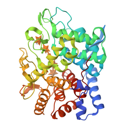
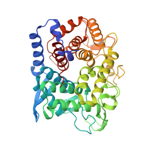
Structure and function of a glycoside hydrolase family 8 endoxylanase from Teredinibacter turnerae.

Fowler, C.A.Hemsworth, G.R.Cuskin, F.Hart, S.Turkenburg, J.Gilbert, H.J.Walton, P.H.Davies, G.J.

(2018) Acta Crystallogr D Struct Biol 74: 946-955

  • DOI: https://doi.org/10.1107/S2059798318009737
  • Primary Citation of Related Structures:  
    6G00, 6G09, 6G0B, 6G0N

  • PubMed Abstract: 

    The biological conversion of lignocellulosic matter into high-value chemicals or biofuels is of increasing industrial importance as the sector slowly transitions away from nonrenewable sources. Many industrial processes involve the use of cellulolytic enzyme cocktails - a selection of glycoside hydrolases and, increasingly, polysaccharide oxygenases - to break down recalcitrant plant polysaccharides. ORFs from the genome of Teredinibacter turnerae, a symbiont hosted within the gills of marine shipworms, were identified in order to search for enzymes with desirable traits. Here, a putative T. turnerae glycoside hydrolase from family 8, hereafter referred to as TtGH8, is analysed. The enzyme is shown to be active against β-1,4-xylan and mixed-linkage (β-1,3,β-1,4) marine xylan. Kinetic parameters, obtained using high-performance anion-exchange chromatography with pulsed amperometric detection and 3,5-dinitrosalicyclic acid reducing-sugar assays, show that TtGH8 catalyses the hydrolysis of β-1,4-xylohexaose with a k cat /K m of 7.5 × 10 7  M -1  min -1 but displays maximal activity against mixed-linkage polymeric xylans, hinting at a primary role in the degradation of marine polysaccharides. The three-dimensional structure of TtGH8 was solved in uncomplexed and xylobiose-, xylotriose- and xylohexaose-bound forms at approximately 1.5 Å resolution; the latter was consistent with the greater k cat /K m for hexasaccharide substrates. A 2,5 B boat conformation observed in the -1 position of bound xylotriose is consistent with the proposed conformational itinerary for this class of enzyme. This work shows TtGH8 to be effective at the degradation of xylan-based substrates, notably marine xylan, further exemplifying the potential of T. turnerae for effective and diverse biomass degradation.


  • Organizational Affiliation

    York Structural Biology Laboratory, Department of Chemistry, The University of York, York YO10 5DD, England.


Macromolecules
Find similar proteins by:  (by identity cutoff)  |  3D Structure
Entity ID: 1
MoleculeChains Sequence LengthOrganismDetailsImage
Glycoside hydrolase family 8 domain protein399Teredinibacter turnerae T7901Mutation(s): 1 
Gene Names: TERTU_4506
EC: 3.2.1 (PDB Primary Data), 3.2.1.4 (UniProt)
UniProt
Find proteins for C5BJ89 (Teredinibacter turnerae (strain ATCC 39867 / T7901))
Explore C5BJ89 
Go to UniProtKB:  C5BJ89
Entity Groups  
Sequence Clusters30% Identity50% Identity70% Identity90% Identity95% Identity100% Identity
UniProt GroupC5BJ89
Sequence Annotations
Expand
  • Reference Sequence
Oligosaccharides

Help

Entity ID: 2
MoleculeChains Length2D Diagram Glycosylation3D Interactions
beta-D-xylopyranose-(1-4)-beta-D-xylopyranose-(1-4)-beta-D-xylopyranose-(1-4)-beta-D-xylopyranose-(1-4)-beta-D-xylopyranose
B
5N/A
Glycosylation Resources
GlyTouCan:  G47101GE
GlyCosmos:  G47101GE
Experimental Data & Validation

Experimental Data

  • Method: X-RAY DIFFRACTION
  • Resolution: 1.80 Å
  • R-Value Free:  0.199 (Depositor), 0.210 (DCC) 
  • R-Value Work:  0.169 (Depositor), 0.180 (DCC) 
  • R-Value Observed: 0.170 (Depositor) 
Space Group: P 21 21 21
Unit Cell:
Length ( Å )Angle ( ˚ )
a = 60.973α = 90
b = 79.694β = 90
c = 88.01γ = 90
Software Package:
Software NamePurpose
REFMACrefinement
iMOSFLMdata reduction
Aimlessdata scaling
MOLREPphasing

Structure Validation

View Full Validation Report


Ligand Structure Quality Assessment 

Created with Raphaël 2.3.0Worse 01 BetterLigand structure goodness of fit to experimental dataBest fitted XYPClick on this verticalbar to view details

Entry History & Funding Information

Deposition Data


Funding OrganizationLocationGrant Number
Biotechnology and Biological Sciences Research CouncilUnited KingdomBB/L001926/1

Revision History  (Full details and data files)

  • Version 1.0: 2018-10-10
    Type: Initial release
  • Version 1.1: 2018-10-17
    Changes: Data collection, Database references
  • Version 2.0: 2020-07-29
    Type: Remediation
    Reason: Carbohydrate remediation
    Changes: Advisory, Atomic model, Data collection, Derived calculations, Structure summary
  • Version 2.1: 2024-05-08
    Changes: Data collection, Database references, Derived calculations, Structure summary