4Z8K | pdb_00004z8k

Phycocyanin structure from T. elongatus at 2.5-A from XFEL using a viscous delivery medium for serial femtosecond crystallography


Experimental Data Snapshot

  • Method: X-RAY DIFFRACTION
  • Resolution: 2.50 Å
  • R-Value Free: 
    0.255 (Depositor), 0.260 (DCC) 
  • R-Value Work: 
    0.187 (Depositor), 0.190 (DCC) 
  • R-Value Observed: 
    0.191 (Depositor) 

Starting Model: experimental
View more details

wwPDB Validation   3D Report Full Report


Ligand Structure Quality Assessment 

Created with Raphaël 2.3.0Worse 01 BetterLigand structure goodness of fit to experimental dataBest fitted CYCClick on this verticalbar to view details

This is version 1.5 of the entry. See complete history


Literature

A novel inert crystal delivery medium for serial femtosecond crystallography.

Conrad, C.E.Basu, S.James, D.Wang, D.Schaffer, A.Roy-Chowdhury, S.Zatsepin, N.A.Aquila, A.Coe, J.Gati, C.Hunter, M.S.Koglin, J.E.Kupitz, C.Nelson, G.Subramanian, G.White, T.A.Zhao, Y.Zook, J.Boutet, S.Cherezov, V.Spence, J.C.Fromme, R.Weierstall, U.Fromme, P.

(2015) IUCrJ 2: 421-430

  • DOI: https://doi.org/10.1107/S2052252515009811
  • Primary Citation of Related Structures:  
    4Z8K

  • PubMed Abstract: 

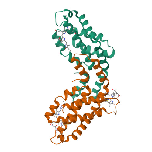
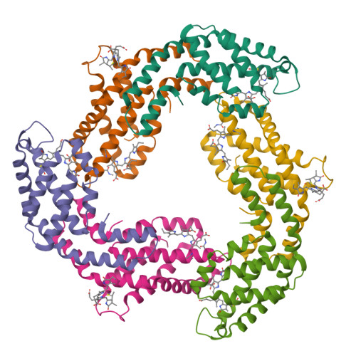
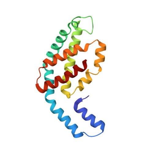
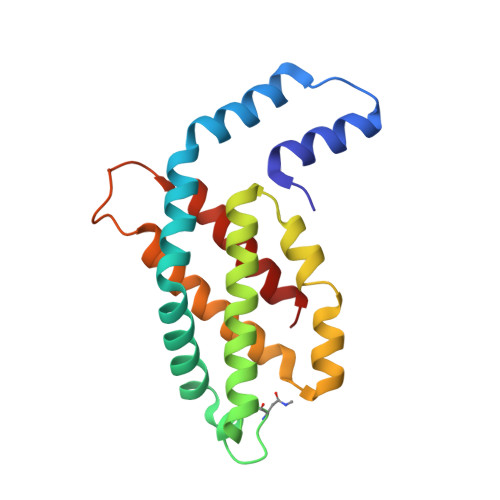
    Serial femtosecond crystallography (SFX) has opened a new era in crystallo-graphy by permitting nearly damage-free, room-temperature structure determination of challenging proteins such as membrane proteins. In SFX, femtosecond X-ray free-electron laser pulses produce diffraction snapshots from nanocrystals and microcrystals delivered in a liquid jet, which leads to high protein consumption. A slow-moving stream of agarose has been developed as a new crystal delivery medium for SFX. It has low background scattering, is compatible with both soluble and membrane proteins, and can deliver the protein crystals at a wide range of temperatures down to 4°C. Using this crystal-laden agarose stream, the structure of a multi-subunit complex, phycocyanin, was solved to 2.5 Å resolution using 300 µg of microcrystals embedded into the agarose medium post-crystallization. The agarose delivery method reduces protein consumption by at least 100-fold and has the potential to be used for a diverse population of proteins, including membrane protein complexes.


  • Organizational Affiliation

    Department of Chemistry and Biochemistry, Arizona State University , PO Box 871604, Tempe, AZ 85287-1604, USA ; Center for Applied Structural Discovery, The Biodesign Institute , PO Box 875001, Tempe, AZ 85287-5001, USA.


Macromolecules
Find similar proteins by:  (by identity cutoff)  |  3D Structure
Entity ID: 1
MoleculeChains Sequence LengthOrganismDetailsImage
C-phycocyanin alpha chain162Thermosynechococcus vestitus BP-1Mutation(s): 0 
UniProt
Find proteins for P50032 (Thermosynechococcus vestitus (strain NIES-2133 / IAM M-273 / BP-1))
Explore P50032 
Go to UniProtKB:  P50032
Entity Groups  
Sequence Clusters30% Identity50% Identity70% Identity90% Identity95% Identity100% Identity
UniProt GroupP50032
Sequence Annotations
Expand
  • Reference Sequence
Find similar proteins by:  (by identity cutoff)  |  3D Structure
Entity ID: 2
MoleculeChains Sequence LengthOrganismDetailsImage
C-phycocyanin beta chain172Thermosynechococcus vestitus BP-1Mutation(s): 0 
UniProt
Find proteins for P50033 (Thermosynechococcus vestitus (strain NIES-2133 / IAM M-273 / BP-1))
Explore P50033 
Go to UniProtKB:  P50033
Entity Groups  
Sequence Clusters30% Identity50% Identity70% Identity90% Identity95% Identity100% Identity
UniProt GroupP50033
Sequence Annotations
Expand
  • Reference Sequence
Experimental Data & Validation

Experimental Data

  • Method: X-RAY DIFFRACTION
  • Resolution: 2.50 Å
  • R-Value Free:  0.255 (Depositor), 0.260 (DCC) 
  • R-Value Work:  0.187 (Depositor), 0.190 (DCC) 
  • R-Value Observed: 0.191 (Depositor) 
Space Group: P 63
Unit Cell:
Length ( Å )Angle ( ˚ )
a = 153.4α = 90
b = 153.4β = 90
c = 39.64γ = 120
Software Package:
Software NamePurpose
PHENIXrefinement
PHASERphasing

Structure Validation

View Full Validation Report


Ligand Structure Quality Assessment 

Created with Raphaël 2.3.0Worse 01 BetterLigand structure goodness of fit to experimental dataBest fitted CYCClick on this verticalbar to view details

Entry History & Funding Information

Deposition Data


Funding OrganizationLocationGrant Number
National Institutes of Health/National Institute of General Medical Sciences (NIH/NIGMS)United StatesR01 GM095583
National Institutes of Health/National Institute of General Medical Sciences (NIH/NIGMS)United StatesU54GM094599
National Institutes of Health/National Institute of General Medical Sciences (NIH/NIGMS)United StatesR01 GM108635
Department of Energy (DOE, United States)United StatesDE-SC001057
National Institutes of Health/National Institute of General Medical Sciences (NIH/NIGMS)United StatesGM097463

Revision History  (Full details and data files)

  • Version 1.0: 2015-08-12
    Type: Initial release
  • Version 1.1: 2018-02-14
    Changes: Author supporting evidence, Data collection, Derived calculations
  • Version 1.2: 2018-09-19
    Changes: Data collection
  • Version 1.3: 2019-12-04
    Changes: Author supporting evidence
  • Version 1.4: 2022-03-23
    Changes: Author supporting evidence, Database references
  • Version 1.5: 2023-09-27
    Changes: Data collection, Refinement description