3VU2 | pdb_00003vu2

Structure of the Starch Branching Enzyme I (BEI) complexed with maltopentaose from Oryza sativa L


Experimental Data Snapshot

  • Method: X-RAY DIFFRACTION
  • Resolution: 2.23 Å
  • R-Value Free: 
    0.286 (Depositor), 0.280 (DCC) 
  • R-Value Work: 
    0.226 (Depositor), 0.220 (DCC) 
  • R-Value Observed: 
    0.229 (Depositor) 

wwPDB Validation   3D Report Full Report


Ligand Structure Quality Assessment 

Created with Raphaël 2.3.0Worse 01 BetterLigand structure goodness of fit to experimental dataBest fitted BGCClick on this verticalbar to view details

This is version 2.1 of the entry. See complete history


Literature

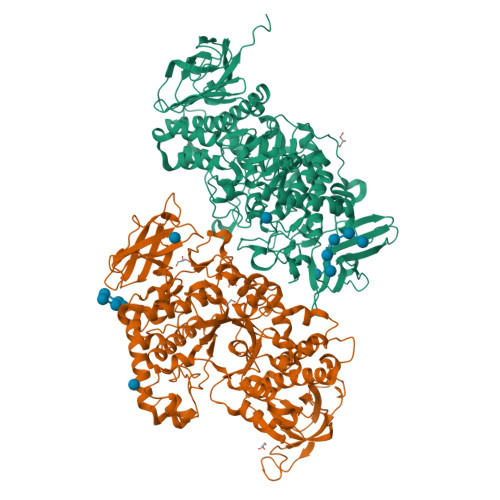
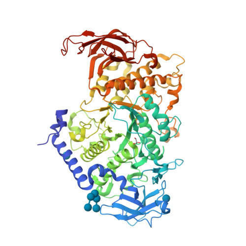
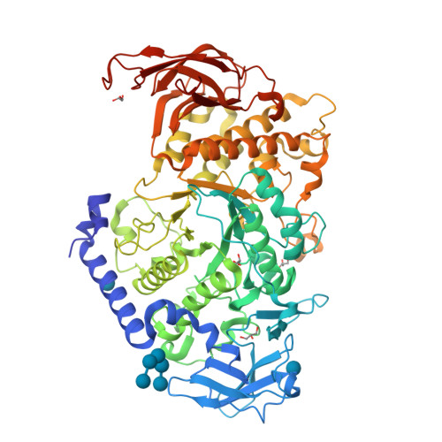
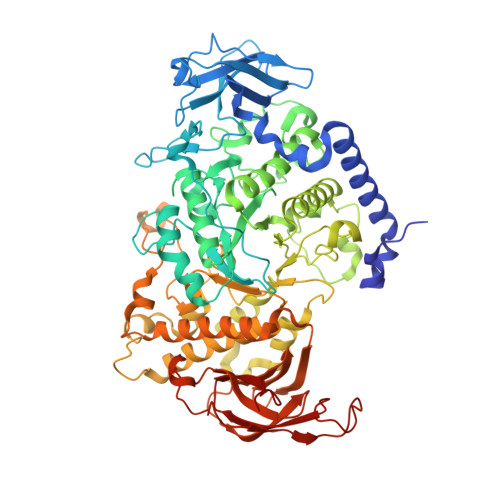
Crystal structure of the rice branching enzyme I (BEI) in complex with maltopentaose.

Chaen, K.Noguchi, J.Omori, T.Kakuta, Y.Kimura, M.

(2012) Biochem Biophys Res Commun 424: 508-511

  • DOI: https://doi.org/10.1016/j.bbrc.2012.06.145
  • Primary Citation of Related Structures:  
    3VU2

  • PubMed Abstract: 

    Starch branching enzyme (SBE) catalyzes the cleavage of α-1,4-linkages and the subsequent transfer of α-1,4 glucan to form an α-1,6 branch point in amylopectin. We determined the crystal structure of the rice branching enzyme I (BEI) in complex with maltopentaose at a resolution of 2.2Å. Maltopentaose bound to a hydrophobic pocket formed by the N-terminal helix, carbohydrate-binding module 48 (CBM48), and α-amylase domain. In addition, glucose moieties could be observed at molecular surfaces on the N-terminal helix (α2) and CBM48. Amino acid residues involved in the carbohydrate bindings are highly conserved in other SBEs, suggesting their generally conserved role in substrate binding for SBEs.


  • Organizational Affiliation

    Laboratory of Structural Biology, Graduate School of Systems Life Sciences, Kyushu University, Hakozaki 6-10-1, Fukuoka 812-8581, Japan.


Macromolecules
Find similar proteins by:  (by identity cutoff)  |  3D Structure
Entity ID: 1
MoleculeChains Sequence LengthOrganismDetailsImage
1,4-alpha-glucan-branching enzyme, chloroplastic/amyloplastic
A, B
702Oryza sativa Japonica GroupMutation(s): 1 
Gene Names: SBE1RBE1Os06g0726400LOC_Os06g51084P0017G10.8-1P0017G10.8-2P0548E04.28-1P0548E04.28-2
EC: 2.4.1.18
UniProt
Find proteins for Q01401 (Oryza sativa subsp. japonica)
Explore Q01401 
Go to UniProtKB:  Q01401
Entity Groups  
Sequence Clusters30% Identity50% Identity70% Identity90% Identity95% Identity100% Identity
UniProt GroupQ01401
Sequence Annotations
Expand
  • Reference Sequence
Oligosaccharides

Help

Entity ID: 2
MoleculeChains Length2D Diagram Glycosylation3D Interactions
alpha-D-glucopyranose-(1-4)-alpha-D-glucopyranose-(1-4)-alpha-D-glucopyranose-(1-4)-alpha-D-glucopyranose-(1-4)-alpha-D-glucopyranose
C, D
5N/A
Glycosylation Resources
GlyTouCan:  G50146AM
GlyCosmos:  G50146AM
Small Molecules
Ligands 2 Unique
IDChains Name / Formula / InChI Key2D Diagram3D Interactions
BGC
Query on BGC

Download Ideal Coordinates CCD File 
E [auth A],
F [auth A],
H [auth B],
I [auth B]
beta-D-glucopyranose
C6 H12 O6
WQZGKKKJIJFFOK-VFUOTHLCSA-N
GOL
Query on GOL

Download Ideal Coordinates CCD File 
G [auth A],
J [auth B],
K [auth B],
L [auth B],
M [auth B]
GLYCEROL
C3 H8 O3
PEDCQBHIVMGVHV-UHFFFAOYSA-N
Biologically Interesting Molecules (External Reference) 1 Unique
Experimental Data & Validation

Experimental Data

  • Method: X-RAY DIFFRACTION
  • Resolution: 2.23 Å
  • R-Value Free:  0.286 (Depositor), 0.280 (DCC) 
  • R-Value Work:  0.226 (Depositor), 0.220 (DCC) 
  • R-Value Observed: 0.229 (Depositor) 
Space Group: P 1 21 1
Unit Cell:
Length ( Å )Angle ( ˚ )
a = 73.81α = 90
b = 66.56β = 89.97
c = 169.625γ = 90
Software Package:
Software NamePurpose
ADSCdata collection
MOLREPphasing
REFMACrefinement
HKL-2000data reduction
HKL-2000data scaling

Structure Validation

View Full Validation Report


Ligand Structure Quality Assessment 

Created with Raphaël 2.3.0Worse 01 BetterLigand structure goodness of fit to experimental dataBest fitted BGCClick on this verticalbar to view details

Entry History 

Deposition Data

Revision History  (Full details and data files)

  • Version 1.0: 2013-05-08
    Type: Initial release
  • Version 2.0: 2020-07-29
    Type: Remediation
    Reason: Carbohydrate remediation
    Changes: Atomic model, Data collection, Database references, Derived calculations, Non-polymer description, Structure summary
  • Version 2.1: 2024-03-20
    Changes: Data collection, Database references, Structure summary