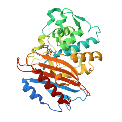
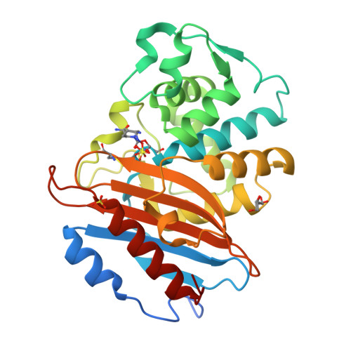
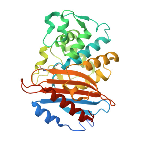
8RWP | pdb_00008rwp

KPC-2 G89D Mutant in Complex with Avibactam


Experimental Data Snapshot

  • Method: X-RAY DIFFRACTION
  • Resolution: 1.19 Å
  • R-Value Free: 
    0.179 (Depositor), 0.188 (DCC) 
  • R-Value Work: 
    0.155 (Depositor), 0.169 (DCC) 
  • R-Value Observed: 
    0.157 (Depositor) 

Starting Model: experimental
View more details

wwPDB Validation   3D Report Full Report


Ligand Structure Quality Assessment 

Created with Raphaël 2.3.0Worse 01 BetterLigand structure goodness of fit to experimental dataBest fitted NXLClick on this verticalbar to view detailsBest fitted A1H3MClick on this verticalbar to view details

This is version 1.0 of the entry. See complete history


Literature

Dynamical responses predict a distal site that modulates activity in an antibiotic resistance enzyme.

Beer, M.Oliveira, A.S.F.Tooke, C.L.Hinchliffe, P.Tsz Yan Li, A.Balega, B.Spencer, J.Mulholland, A.J.

(2024) Chem Sci 15: 17232-17244

  • DOI: https://doi.org/10.1039/d4sc03295k
  • Primary Citation of Related Structures:  
    8RWO, 8RWP, 8RWQ, 8RWR, 8RWS

  • PubMed Abstract: 

    β-Lactamases, which hydrolyse β-lactam antibiotics, are key determinants of antibiotic resistance. Predicting the sites and effects of distal mutations in enzymes is challenging. For β-lactamases, the ability to make such predictions would contribute to understanding activity against, and development of, antibiotics and inhibitors to combat resistance. Here, using dynamical non-equilibrium molecular dynamics (D-NEMD) simulations combined with experiments, we demonstrate that intramolecular communication networks differ in three class A SulpHydryl Variant (SHV)-type β-lactamases. Differences in network architecture and correlated motions link to catalytic efficiency and β-lactam substrate spectrum. Further, the simulations identify a distal residue at position 89 in the clinically important Klebsiella pneumoniae carbapenemase 2 (KPC-2), as a participant in similar networks, suggesting that mutation at this position would modulate enzyme activity. Experimental kinetic, biophysical and structural characterisation of the naturally occurring, but previously biochemically uncharacterised, KPC-2 G89D mutant with several antibiotics and inhibitors reveals significant changes in hydrolytic spectrum, specifically reducing activity towards carbapenems without effecting major structural or stability changes. These results show that D-NEMD simulations can predict distal sites where mutation affects enzyme activity. This approach could have broad application in understanding enzyme evolution, and in engineering of natural and de novo enzymes.


  • Organizational Affiliation

    School of Cellular and Molecular Medicine, University of Bristol Bristol BS8 1TD UK Jim.spencer@bristol.ac.uk.


Macromolecules
Find similar proteins by:  (by identity cutoff)  |  3D Structure
Entity ID: 1
MoleculeChains Sequence LengthOrganismDetailsImage
Carbapenem-hydrolyzing beta-lactamase KPC290Klebsiella pneumoniaeMutation(s): 1 
Gene Names: blakpckpc1
EC: 3.5.2.6
UniProt
Find proteins for Q9F663 (Klebsiella pneumoniae)
Explore Q9F663 
Go to UniProtKB:  Q9F663
Entity Groups  
Sequence Clusters30% Identity50% Identity70% Identity90% Identity95% Identity100% Identity
UniProt GroupQ9F663
Sequence Annotations
Expand
  • Reference Sequence
Small Molecules
Ligands 4 Unique
IDChains Name / Formula / InChI Key2D Diagram3D Interactions
NXL (Subject of Investigation/LOI)
Query on NXL

Download Ideal Coordinates CCD File 
C [auth A](2S,5R)-1-formyl-5-[(sulfooxy)amino]piperidine-2-carboxamide
C7 H13 N3 O6 S
WJDGWXPPFHLLNL-RITPCOANSA-N
A1H3M (Subject of Investigation/LOI)
Query on A1H3M

Download Ideal Coordinates CCD File 
D [auth A](2~{S})-5-azanylidene-1-methanoyl-piperidine-2-carboxamide
C7 H11 N3 O2
SVUBAXFAVRYYJU-YTDSJMIDSA-N
SO4
Query on SO4

Download Ideal Coordinates CCD File 
E [auth A],
F [auth A]
SULFATE ION
O4 S
QAOWNCQODCNURD-UHFFFAOYSA-L
GOL
Query on GOL

Download Ideal Coordinates CCD File 
B [auth A]GLYCEROL
C3 H8 O3
PEDCQBHIVMGVHV-UHFFFAOYSA-N
Experimental Data & Validation

Experimental Data

  • Method: X-RAY DIFFRACTION
  • Resolution: 1.19 Å
  • R-Value Free:  0.179 (Depositor), 0.188 (DCC) 
  • R-Value Work:  0.155 (Depositor), 0.169 (DCC) 
  • R-Value Observed: 0.157 (Depositor) 
Space Group: P 21 21 2
Unit Cell:
Length ( Å )Angle ( ˚ )
a = 60.14α = 90
b = 78.49β = 90
c = 55.64γ = 90
Software Package:
Software NamePurpose
PHENIXrefinement
DIALSdata reduction
DIALSdata scaling
PHASERphasing

Structure Validation

View Full Validation Report


Ligand Structure Quality Assessment 

Created with Raphaël 2.3.0Worse 01 BetterLigand structure goodness of fit to experimental dataBest fitted NXLClick on this verticalbar to view detailsBest fitted A1H3MClick on this verticalbar to view details

Entry History & Funding Information

Deposition Data


Funding OrganizationLocationGrant Number
Biotechnology and Biological Sciences Research Council (BBSRC)United KingdomBB/T008741/10
European Research Council (ERC)European Union101021207

Revision History  (Full details and data files)

  • Version 1.0: 2025-01-22
    Type: Initial release