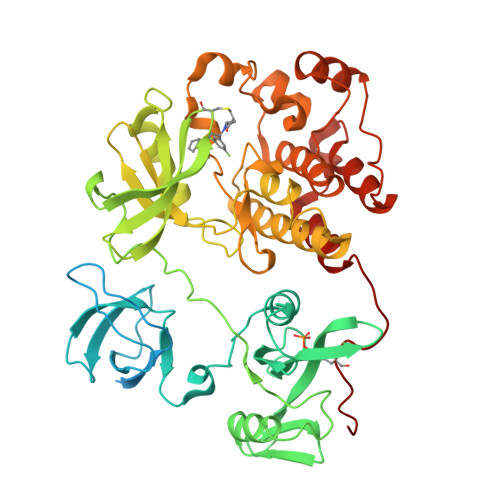
The crystal structure of c-Src in complex with covalent inhibitor LW-Srci-8
Zhang, H.M., Luo, C.To be published.
Experimental Data Snapshot
Starting Model: experimental
View more details
Entity ID: 1 | |||||
|---|---|---|---|---|---|
| Molecule | Chains | Sequence Length | Organism | Details | Image |
| Proto-oncogene tyrosine-protein kinase Src | 536 | Homo sapiens | Mutation(s): 0 Gene Names: SRC, SRC1 EC: 2.7.10.2 |  | |
UniProt & NIH Common Fund Data Resources | |||||
Find proteins for P12931 (Homo sapiens) Explore P12931 Go to UniProtKB: P12931 | |||||
PHAROS: P12931 GTEx: ENSG00000197122 | |||||
Entity Groups | |||||
| Sequence Clusters | 30% Identity50% Identity70% Identity90% Identity95% Identity100% Identity | ||||
| UniProt Group | P12931 | ||||
Sequence AnnotationsExpand | |||||
| |||||
| Ligands 1 Unique | |||||
|---|---|---|---|---|---|
| ID | Chains | Name / Formula / InChI Key | 2D Diagram | 3D Interactions | |
| UJ0 (Subject of Investigation/LOI) Query on UJ0 | B [auth A] | N-[(1R)-1-[3,5-bis(fluoranyl)phenyl]-2-(cyclopentylamino)-2-oxidanylidene-ethyl]-N-cyclopropyl-prop-2-enamide C19 H22 F2 N2 O2 VMRIFQHBFDXKML-GOSISDBHSA-N |  | ||
| Modified Residues 1 Unique | |||||
|---|---|---|---|---|---|
| ID | Chains | Type | Formula | 2D Diagram | Parent |
| PTR Query on PTR | A | L-PEPTIDE LINKING | C9 H12 N O6 P |  | TYR |
| Length ( Å ) | Angle ( ˚ ) |
|---|---|
| a = 51.012 | α = 90 |
| b = 83.11 | β = 90 |
| c = 106.861 | γ = 90 |
| Software Name | Purpose |
|---|---|
| PHENIX | refinement |
| XDS | data reduction |
| XDS | data scaling |
| PHENIX | phasing |
| Funding Organization | Location | Grant Number |
|---|---|---|
| National Natural Science Foundation of China (NSFC) | China | -- |