7P6T

Crystal structure of the FimH-binding decoy module of human glycoprotein 2 (GP2) (crystal form III)


Experimental Data Snapshot

  • Method: X-RAY DIFFRACTION
  • Resolution: 1.40 Å
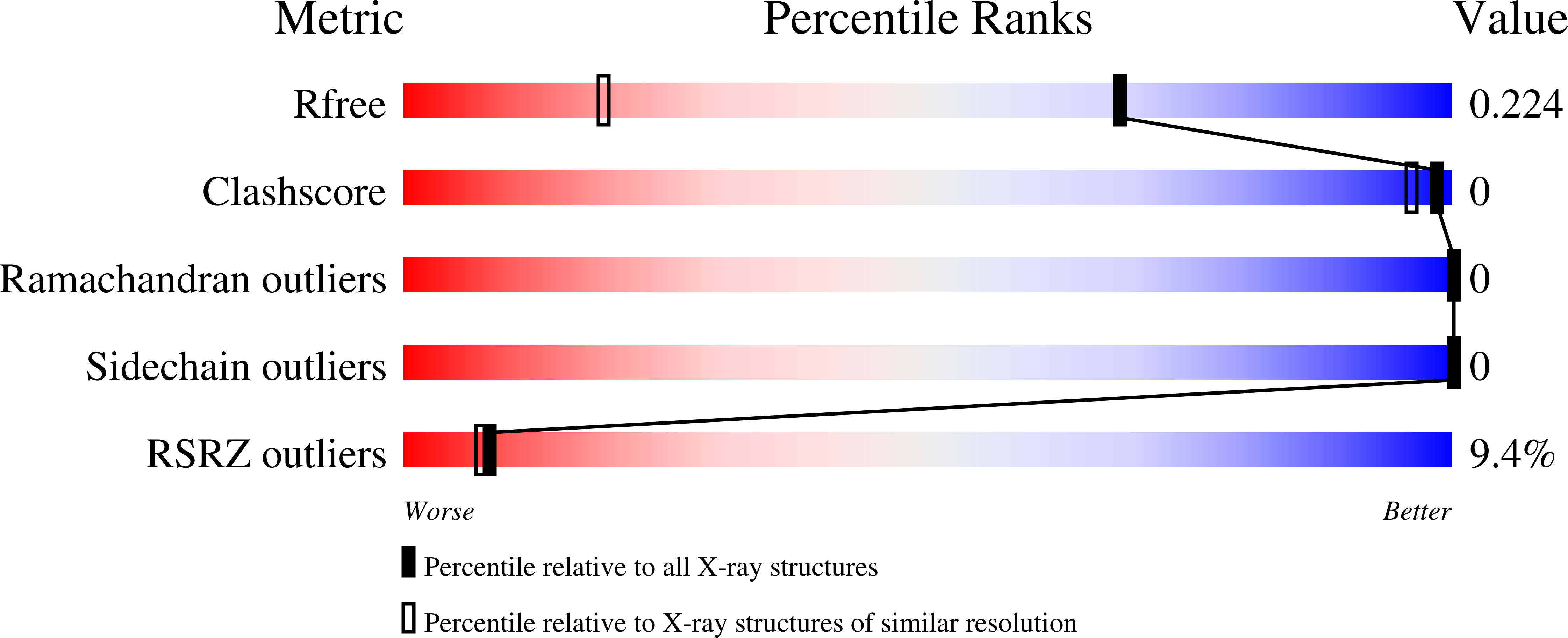
  • R-Value Free: 0.224 
  • R-Value Work: 0.195 
  • R-Value Observed: 0.197 

Starting Model: experimental
View more details

wwPDB Validation   3D Report Full Report


Ligand Structure Quality Assessment 


This is version 1.4 of the entry. See complete history


Literature

Structure of the decoy module of human glycoprotein 2 and uromodulin and its interaction with bacterial adhesin FimH.

Stsiapanava, A.Xu, C.Nishio, S.Han, L.Yamakawa, N.Carroni, M.Tunyasuvunakool, K.Jumper, J.de Sanctis, D.Wu, B.Jovine, L.

(2022) Nat Struct Mol Biol 29: 190-193

  • DOI: https://doi.org/10.1038/s41594-022-00729-3
  • Primary Citation of Related Structures:  
    7P6R, 7P6S, 7P6T, 7PFP, 7Q3N

  • PubMed Abstract: 

    Glycoprotein 2 (GP2) and uromodulin (UMOD) filaments protect against gastrointestinal and urinary tract infections by acting as decoys for bacterial fimbrial lectin FimH. By combining AlphaFold2 predictions with X-ray crystallography and cryo-EM, we show that these proteins contain a bipartite decoy module whose new fold presents the high-mannose glycan recognized by FimH. The structure rationalizes UMOD mutations associated with kidney diseases and visualizes a key epitope implicated in cast nephropathy.


  • Organizational Affiliation

    Department of Biosciences and Nutrition, Karolinska Institutet, Huddinge, Sweden.


Macromolecules
Find similar proteins by:  (by identity cutoff)  |  3D Structure
Entity ID: 1
MoleculeChains Sequence LengthOrganismDetailsImage
Isoform Alpha of Pancreatic secretory granule membrane major glycoprotein GP2161Homo sapiensMutation(s): 0 
Gene Names: GP2
UniProt & NIH Common Fund Data Resources
Find proteins for P55259 (Homo sapiens)
Explore P55259 
Go to UniProtKB:  P55259
PHAROS:  P55259
GTEx:  ENSG00000169347 
Entity Groups  
Sequence Clusters30% Identity50% Identity70% Identity90% Identity95% Identity100% Identity
UniProt GroupP55259
Glycosylation
Glycosylation Sites: 3Go to GlyGen: P55259-1
Sequence Annotations
Expand
  • Reference Sequence
Small Molecules
Ligands 2 Unique
IDChains Name / Formula / InChI Key2D Diagram3D Interactions
NAG (Subject of Investigation/LOI)
Query on NAG

Download Ideal Coordinates CCD File 
B [auth A],
C [auth A],
D [auth A]
2-acetamido-2-deoxy-beta-D-glucopyranose
C8 H15 N O6
OVRNDRQMDRJTHS-FMDGEEDCSA-N
9D2
Query on 9D2

Download Ideal Coordinates CCD File 
E [auth A],
F [auth A],
G [auth A],
H [auth A],
I [auth A]
2-ethyl-2-(hydroxymethyl)propane-1,3-diol
C6 H14 O3
ZJCCRDAZUWHFQH-UHFFFAOYSA-N
Experimental Data & Validation

Experimental Data

  • Method: X-RAY DIFFRACTION
  • Resolution: 1.40 Å
  • R-Value Free: 0.224 
  • R-Value Work: 0.195 
  • R-Value Observed: 0.197 
  • Space Group: C 1 2 1
Unit Cell:
Length ( Å )Angle ( ˚ )
a = 90.15α = 90
b = 33.66β = 111.839
c = 59.63γ = 90
Software Package:
Software NamePurpose
MxCuBEdata collection
XDSdata reduction
XSCALEdata scaling
PHASERphasing
Cootmodel building
ISOLDErefinement
PHENIXrefinement

Structure Validation

View Full Validation Report


Ligand Structure Quality Assessment 


Entry History & Funding Information

Deposition Data


Funding OrganizationLocationGrant Number
Swedish Research CouncilSweden2016-03999
Swedish Research CouncilSweden2020-04936
Knut and Alice Wallenberg FoundationSweden2018.0042

Revision History  (Full details and data files)

  • Version 1.0: 2022-03-16
    Type: Initial release
  • Version 1.1: 2022-03-23
    Changes: Database references
  • Version 1.2: 2022-03-30
    Changes: Database references
  • Version 1.3: 2024-05-01
    Changes: Data collection, Refinement description
  • Version 1.4: 2024-10-09
    Changes: Structure summary