7NA2

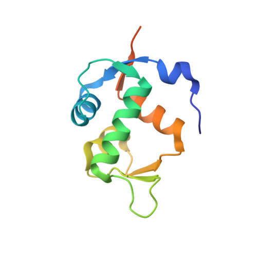
HDM2 in complex with compound 56


Experimental Data Snapshot

  • Method: X-RAY DIFFRACTION
  • Resolution: 1.86 Å
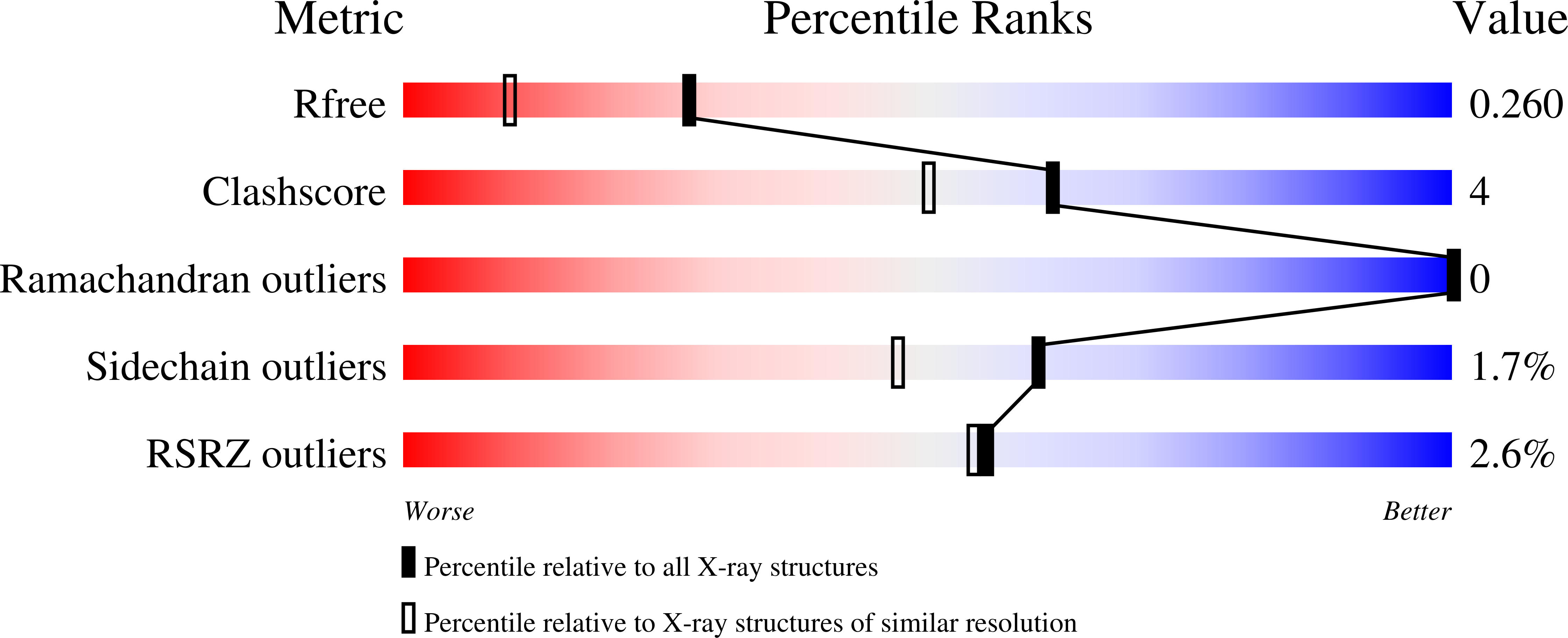
  • R-Value Free: 0.259 
  • R-Value Work: 0.186 
  • R-Value Observed: 0.193 

wwPDB Validation   3D Report Full Report


Ligand Structure Quality Assessment 


This is version 1.2 of the entry. See complete history


Literature

Discovery of MK-4688 : an Efficient Inhibitor of the HDM2-p53 Protein-Protein Interaction.

Reutershan, M.H.Machacek, M.R.Altman, M.D.Bogen, S.Cai, M.Cammarano, C.Chen, D.Christopher, M.Cryan, J.Daublain, P.Fradera, X.Geda, P.Goldenblatt, P.Hill, A.D.Kemper, R.A.Kutilek, V.Li, C.Martinez, M.McCoy, M.Nair, L.Pan, W.Thompson, C.F.Scapin, G.Shizuka, M.Spatz, M.L.Steinhuebel, D.Sun, B.Voss, M.E.Wang, X.Yang, L.Yeh, T.C.Dussault, I.Marshall, C.G.Trotter, B.W.

(2021) J Med Chem 64: 16213-16241

  • DOI: https://doi.org/10.1021/acs.jmedchem.1c01524
  • Primary Citation of Related Structures:  
    7NA1, 7NA2, 7NA3, 7NA4

  • PubMed Abstract: 

    Identification of low-dose, low-molecular-weight, drug-like inhibitors of protein-protein interactions (PPIs) is a challenging area of research. Despite the challenges, the therapeutic potential of PPI inhibition has driven significant efforts toward this goal. Adding to recent success in this area, we describe herein our efforts to optimize a novel purine carboxylic acid-derived inhibitor of the HDM2-p53 PPI into a series of low-projected dose inhibitors with overall favorable pharmacokinetic and physical properties. Ultimately, a strategy focused on leveraging known binding hot spots coupled with biostructural information to guide the design of conformationally constrained analogs and a focus on efficiency metrics led to the discovery of MK-4688 (compound 56 ), a highly potent, selective, and low-molecular-weight inhibitor suitable for clinical investigation.


  • Organizational Affiliation

    Merck & Co., Inc., 33 Avenue Louis Pasteur, Boston, Massachusetts 02115, United States.


Macromolecules
Find similar proteins by:  (by identity cutoff)  |  3D Structure
Entity ID: 1
MoleculeChains Sequence LengthOrganismDetailsImage
Isoform 11 of E3 ubiquitin-protein ligase Mdm2
A, B
110Homo sapiensMutation(s): 0 
Gene Names: MDM2
EC: 2.3.2.27
UniProt & NIH Common Fund Data Resources
Find proteins for Q00987 (Homo sapiens)
Explore Q00987 
Go to UniProtKB:  Q00987
PHAROS:  Q00987
GTEx:  ENSG00000135679 
Entity Groups  
Sequence Clusters30% Identity50% Identity70% Identity90% Identity95% Identity100% Identity
UniProt GroupQ00987
Sequence Annotations
Expand
  • Reference Sequence
Small Molecules
Ligands 1 Unique
IDChains Name / Formula / InChI Key2D Diagram3D Interactions
1I0 (Subject of Investigation/LOI)
Query on 1I0

Download Ideal Coordinates CCD File 
C [auth A],
D [auth B]
3-[4-(5-chloropyridin-3-yl)-2-[(4aR,7aR)-hexahydrocyclopenta[b][1,4]oxazin-4(4aH)-yl]-3-{[(1r,4R)-4-methylcyclohexyl]methyl}-3H-imidazo[4,5-c]pyridin-6-yl]-1,2,4-oxadiazol-5(4H)-one
C28 H32 Cl N7 O3
FDBWTMMKIZGUEZ-IEGYKGNPSA-N
Binding Affinity Annotations 
IDSourceBinding Affinity
1I0 BindingDB:  7NA2 IC50: 1 (nM) from 1 assay(s)
Experimental Data & Validation

Experimental Data

  • Method: X-RAY DIFFRACTION
  • Resolution: 1.86 Å
  • R-Value Free: 0.259 
  • R-Value Work: 0.186 
  • R-Value Observed: 0.193 
  • Space Group: P 1 21 1
Unit Cell:
Length ( Å )Angle ( ˚ )
a = 37.925α = 90
b = 68.341β = 113.12
c = 42.105γ = 90
Software Package:
Software NamePurpose
XSCALEdata scaling
REFMACrefinement
PDB_EXTRACTdata extraction
XDSdata reduction
REFMACphasing

Structure Validation

View Full Validation Report


Ligand Structure Quality Assessment 


Entry History 

Deposition Data

  • Released Date: 2021-11-10 
  • Deposition Author(s): Scapin, G.

Revision History  (Full details and data files)

  • Version 1.0: 2021-11-10
    Type: Initial release
  • Version 1.1: 2021-11-24
    Changes: Database references
  • Version 1.2: 2024-05-22
    Changes: Data collection