6W7E

K2P2.1 (TREK-1), 30 mM K+


Experimental Data Snapshot

  • Method: X-RAY DIFFRACTION
  • Resolution: 3.29 Å
  • R-Value Free: 0.307 
  • R-Value Work: 0.288 
  • R-Value Observed: 0.289 

Starting Model: experimental
View more details

wwPDB Validation   3D Report Full Report


Ligand Structure Quality Assessment 

Created with Raphaël 2.3.0Worse 01 BetterLigand structure goodness of fit to experimental dataBest fitted PIOClick on this verticalbar to view detailsBest fitted R16Click on this verticalbar to view detailsBest fitted D12Click on this verticalbar to view details

This is version 1.2 of the entry. See complete history


Literature

K 2P channel C-type gating involves asymmetric selectivity filter order-disorder transitions.

Lolicato, M.Natale, A.M.Abderemane-Ali, F.Crottes, D.Capponi, S.Duman, R.Wagner, A.Rosenberg, J.M.Grabe, M.Minor Jr., D.L.

(2020) Sci Adv 6

  • DOI: https://doi.org/10.1126/sciadv.abc9174
  • Primary Citation of Related Structures:  
    6W7B, 6W7C, 6W7D, 6W7E, 6W82, 6W83, 6W84, 6W85, 6W86, 6W87, 6W88, 6W8A, 6W8C, 6W8F

  • PubMed Abstract: 

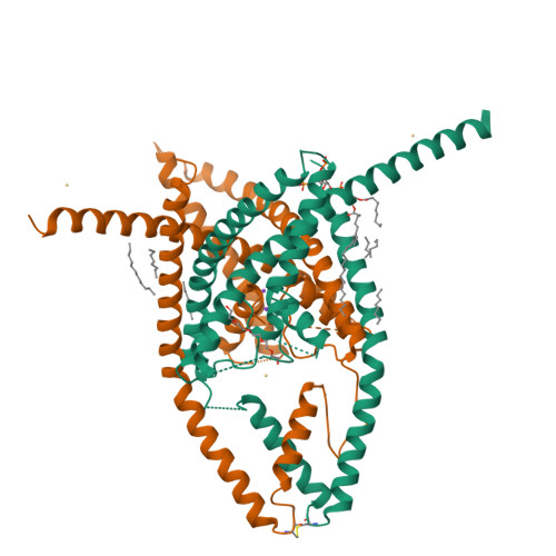
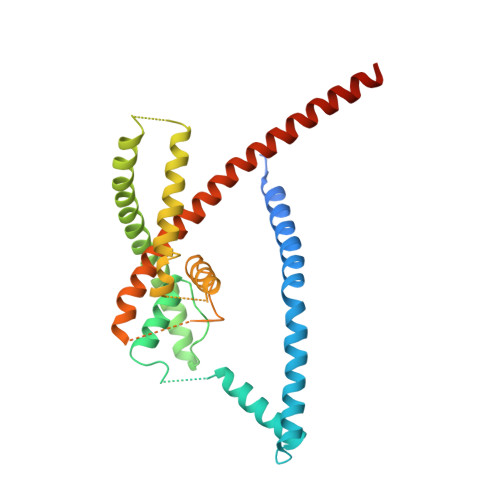
    K 2P potassium channels regulate cellular excitability using their selectivity filter (C-type) gate. C-type gating mechanisms, best characterized in homotetrameric potassium channels, remain controversial and are attributed to selectivity filter pinching, dilation, or subtle structural changes. The extent to which such mechanisms control C-type gating of innately heterodimeric K 2P s is unknown. Here, combining K 2P 2.1 (TREK-1) x-ray crystallography in different potassium concentrations, potassium anomalous scattering, molecular dynamics, and electrophysiology, we uncover unprecedented, asymmetric, potassium-dependent conformational changes that underlie K 2P C-type gating. These asymmetric order-disorder transitions, enabled by the K 2P heterodimeric architecture, encompass pinching and dilation, disrupt the S1 and S2 ion binding sites, require the uniquely long K 2P SF2-M4 loop and conserved "M3 glutamate network," and are suppressed by the K 2P C-type gate activator ML335. These findings demonstrate that two distinct C-type gating mechanisms can operate in one channel and underscore the SF2-M4 loop as a target for K 2P channel modulator development.


  • Organizational Affiliation

    Cardiovascular Research Institute, University of California, San Francisco, CA 93858-2330, USA.


Macromolecules
Find similar proteins by:  (by identity cutoff)  |  3D Structure
Entity ID: 1
MoleculeChains Sequence LengthOrganismDetailsImage
Potassium channel subfamily K member 2
A, B
312Mus musculusMutation(s): 0 
Gene Names: Kcnk2
Membrane Entity: Yes 
UniProt & NIH Common Fund Data Resources
Find proteins for P97438 (Mus musculus)
Explore P97438 
Go to UniProtKB:  P97438
IMPC:  MGI:109366
Entity Groups  
Sequence Clusters30% Identity50% Identity70% Identity90% Identity95% Identity100% Identity
UniProt GroupP97438
Sequence Annotations
Expand
  • Reference Sequence
Small Molecules
Ligands 8 Unique
IDChains Name / Formula / InChI Key2D Diagram3D Interactions
PIO
Query on PIO

Download Ideal Coordinates CCD File 
I [auth A][(2R)-2-octanoyloxy-3-[oxidanyl-[(1R,2R,3S,4R,5R,6S)-2,3,6-tris(oxidanyl)-4,5-diphosphonooxy-cyclohexyl]oxy-phosphoryl]oxy-propyl] octanoate
C25 H49 O19 P3
XLNCEHRXXWQMPK-MJUMVPIBSA-N
1PE
Query on 1PE

Download Ideal Coordinates CCD File 
E [auth A]PENTAETHYLENE GLYCOL
C10 H22 O6
JLFNLZLINWHATN-UHFFFAOYSA-N
R16
Query on R16

Download Ideal Coordinates CCD File 
Q [auth B]HEXADECANE
C16 H34
DCAYPVUWAIABOU-UHFFFAOYSA-N
D12
Query on D12

Download Ideal Coordinates CCD File 
O [auth B]DODECANE
C12 H26
SNRUBQQJIBEYMU-UHFFFAOYSA-N
OCT
Query on OCT

Download Ideal Coordinates CCD File 
G [auth A],
M [auth B],
N [auth B]
N-OCTANE
C8 H18
TVMXDCGIABBOFY-UHFFFAOYSA-N
CD
Query on CD

Download Ideal Coordinates CCD File 
C [auth A],
D [auth A],
J [auth B],
K [auth B]
CADMIUM ION
Cd
WLZRMCYVCSSEQC-UHFFFAOYSA-N
LNK
Query on LNK

Download Ideal Coordinates CCD File 
F [auth A],
L [auth B]
PENTANE
C5 H12
OFBQJSOFQDEBGM-UHFFFAOYSA-N
K
Query on K

Download Ideal Coordinates CCD File 
H [auth A],
P [auth B]
POTASSIUM ION
K
NPYPAHLBTDXSSS-UHFFFAOYSA-N
Experimental Data & Validation

Experimental Data

  • Method: X-RAY DIFFRACTION
  • Resolution: 3.29 Å
  • R-Value Free: 0.307 
  • R-Value Work: 0.288 
  • R-Value Observed: 0.289 
  • Space Group: P 21 21 21
Unit Cell:
Length ( Å )Angle ( ˚ )
a = 67.368α = 90
b = 122.534β = 90
c = 126.552γ = 90
Software Package:
Software NamePurpose
BUSTERrefinement
XDSdata reduction
Cootmodel building
PHASERphasing
Aimlessdata processing
Aimlessdata scaling

Structure Validation

View Full Validation Report


Ligand Structure Quality Assessment 

Created with Raphaël 2.3.0Worse 01 BetterLigand structure goodness of fit to experimental dataBest fitted PIOClick on this verticalbar to view detailsBest fitted R16Click on this verticalbar to view detailsBest fitted D12Click on this verticalbar to view details

Entry History & Funding Information

Deposition Data


Funding OrganizationLocationGrant Number
National Institutes of Health/National Institute of Mental Health (NIH/NIMH)United StatesNIH-R01-MH093603

Revision History  (Full details and data files)

  • Version 1.0: 2021-01-27
    Type: Initial release
  • Version 1.1: 2023-10-18
    Changes: Data collection, Database references, Refinement description
  • Version 1.2: 2024-11-13
    Changes: Structure summary