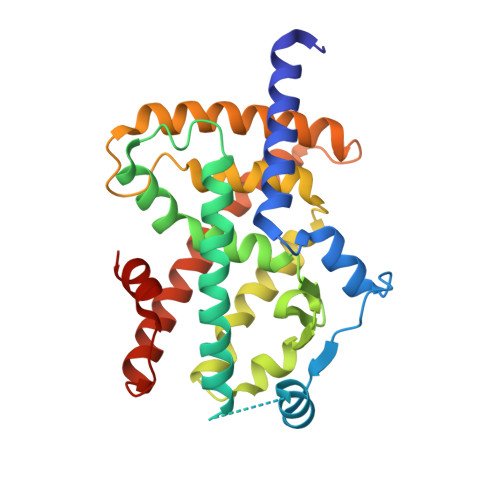
6TSG

Crystal structure of Peroxisome proliferator-activated receptor gamma (PPARG) in complex with TETRAC


Experimental Data Snapshot

  • Method: X-RAY DIFFRACTION
  • Resolution: 2.98 Å
  • R-Value Free: 0.292 
  • R-Value Work: 0.236 
  • R-Value Observed: 0.239 

Starting Model: experimental
View more details

wwPDB Validation   3D Report Full Report


Ligand Structure Quality Assessment 


This is version 1.1 of the entry. See complete history


Literature

l-Thyroxin and the Nonclassical Thyroid Hormone TETRAC Are Potent Activators of PPAR gamma.

Gellrich, L.Heitel, P.Heering, J.Kilu, W.Pollinger, J.Goebel, T.Kahnt, A.Arifi, S.Pogoda, W.Paulke, A.Steinhilber, D.Proschak, E.Wurglics, M.Schubert-Zsilavecz, M.Chaikuad, A.Knapp, S.Bischoff, I.Furst, R.Merk, D.

(2020) J Med Chem 63: 6727-6740

  • DOI: https://doi.org/10.1021/acs.jmedchem.9b02150
  • Primary Citation of Related Structures:  
    6TSG

  • PubMed Abstract: 

    Thyroid hormones (THs) operate numerous physiological processes through modulation of the nuclear thyroid hormone receptors and several other proteins. We report direct activation of the nuclear peroxisome proliferator-activated receptor gamma (PPARγ) and retinoid X receptor (RXR) by classical and nonclassical THs as another molecular activity of THs. The T4 metabolite TETRAC was the most active TH on PPARγ with nanomolar potency and binding affinity. We demonstrate that TETRAC promotes PPARγ/RXR signaling in cell-free, cellular, and in vivo settings. Simultaneous activation of the heterodimer partners PPARγ and RXR resulted in high dimer activation efficacy. Compared to fatty acids as known natural ligands of PPARγ and RXR, TETRAC differs markedly in its molecular structure and the PPARγ-TETRAC complex revealed a distinctive binding mode of the TH. Our observations suggest a potential connection of TH and PPAR signaling through overlapping ligand recognition and may hold implications for TH and PPAR pharmacology.


  • Organizational Affiliation

    Institute of Pharmaceutical Chemistry, Goethe University Frankfurt, Max-von-Laue-Str. 9, D-60438 Frankfurt, Germany.


Macromolecules
Find similar proteins by:  (by identity cutoff)  |  3D Structure
Entity ID: 1
MoleculeChains Sequence LengthOrganismDetailsImage
Peroxisome proliferator-activated receptor gamma277Homo sapiensMutation(s): 0 
Gene Names: PPARGNR1C3
UniProt & NIH Common Fund Data Resources
Find proteins for P37231 (Homo sapiens)
Explore P37231 
Go to UniProtKB:  P37231
PHAROS:  P37231
GTEx:  ENSG00000132170 
Entity Groups  
Sequence Clusters30% Identity50% Identity70% Identity90% Identity95% Identity100% Identity
UniProt GroupP37231
Sequence Annotations
Expand
  • Reference Sequence
Small Molecules
Ligands 1 Unique
IDChains Name / Formula / InChI Key2D Diagram3D Interactions
T4A (Subject of Investigation/LOI)
Query on T4A

Download Ideal Coordinates CCD File 
B [auth A]3,3',5,5'-TETRAIODOTHYROACETIC ACID
C14 H8 I4 O4
PPJYSSNKSXAVDB-UHFFFAOYSA-N
Binding Affinity Annotations 
IDSourceBinding Affinity
T4A BindingDB:  6TSG Kd: 110 (nM) from 1 assay(s)
EC50: 100 (nM) from 1 assay(s)
Experimental Data & Validation

Experimental Data

  • Method: X-RAY DIFFRACTION
  • Resolution: 2.98 Å
  • R-Value Free: 0.292 
  • R-Value Work: 0.236 
  • R-Value Observed: 0.239 
  • Space Group: P 41 21 2
Unit Cell:
Length ( Å )Angle ( ˚ )
a = 61.678α = 90
b = 61.678β = 90
c = 166.487γ = 90
Software Package:
Software NamePurpose
Aimlessdata scaling
REFMACrefinement
PDB_EXTRACTdata extraction
XDSdata reduction
PHASERphasing

Structure Validation

View Full Validation Report


Ligand Structure Quality Assessment 


Entry History 

Deposition Data

Revision History  (Full details and data files)

  • Version 1.0: 2020-07-22
    Type: Initial release
  • Version 1.1: 2024-01-24
    Changes: Data collection, Database references, Refinement description