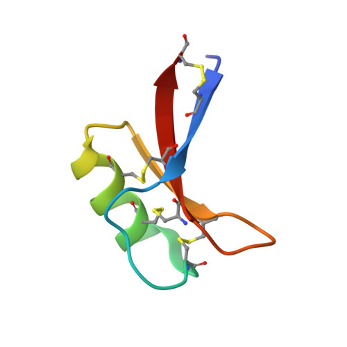
6MRY

NoD173 plant defensin


Experimental Data Snapshot

  • Method: X-RAY DIFFRACTION
  • Resolution: 2.30 Å
  • R-Value Free: 0.232 
  • R-Value Work: 0.189 
  • R-Value Observed: 0.192 

Starting Model: experimental
View more details

wwPDB Validation   3D Report Full Report


Ligand Structure Quality Assessment 


This is version 1.4 of the entry. See complete history


Literature

Structural and functional characterization of the membrane-permeabilizing activity ofNicotiana occidentalisdefensin NoD173 and protein engineering to enhance oncolysis.

Lay, F.T.Ryan, G.F.Caria, S.Phan, T.K.Veneer, P.K.White, J.A.Kvansakul, M.Hulett, M.D.

(2019) FASEB J 33: 6470-6482

  • DOI: https://doi.org/10.1096/fj.201802540R
  • Primary Citation of Related Structures:  
    6MRY

  • PubMed Abstract: 

    Defensins are an extensive family of host defense peptides found ubiquitously across plant and animal species. In addition to protecting against infection by pathogenic microorganisms, some defensins are selectively cytotoxic toward tumor cells. As such, defensins have attracted interest as potential antimicrobial and anticancer therapeutics. The mechanism of defensin action against microbes and tumor cells appears to be conserved and involves the targeting and disruption of cellular membranes. This has been best defined for plant defensins, which upon binding specific phospholipids, such as phosphatidylinositol 4,5-bisphosphate (PIP2) and phosphatidic acid, form defensin-lipid oligomeric complexes that destabilize membranes, leading to cell lysis. In this study, to further define the anticancer and therapeutic properties of plant defensins, we have characterized a novel plant defensin, Nicotiana occidentalis defensin 173 (NoD173), from N. occidentalis. NoD173 at low micromolar concentrations selectively killed a panel of tumor cell lines over normal primary cells. To improve the anticancer activity of NoD173, we explored increasing cationicity by mutation, with NoD173 with the substitution of Q22 with lysine [NoD173(Q22K)], increasing the antitumor cell activity by 2-fold. NoD173 and the NoD173(Q22K) mutant exhibited only low levels of hemolytic activity, and both maintained activity against tumor cells in serum. The ability of NoD173 to inhibit solid tumor growth in vivo was tested in a mouse B16-F1 model, whereby injection of NoD173 into established subcutaneous tumors significantly inhibited tumor growth. Finally, we showed that NoD173 specifically targets PIP2 and determined by X-ray crystallography that a high-resolution structure of NoD173, which forms a conserved family-defining cysteine-stabilized-αβ motif with a dimeric lipid-binding conformation, configured into an arch-shaped oligomer of 4 dimers. These data provide insights into the mechanism of how defensins target membranes to kill tumor cells and provide proof of concept that defensins are able to inhibit tumor growth in vivo .-Lay, F. T., Ryan, G. F., Caria, S., Phan, T. K., Veneer, P. K., White, J. A., Kvansakul, M., Hulett M. D. Structural and functional characterization of the membrane-permeabilizing activity of Nicotiana occidentalis defensin NoD173 and protein engineering to enhance oncolysis.


  • Organizational Affiliation

    Department of Biochemistry and Genetics, La Trobe Institute for Molecular Science, La Trobe University, Melbourne, Victoria, Australia.


Macromolecules
Find similar proteins by:  (by identity cutoff)  |  3D Structure
Entity ID: 1
MoleculeChains Sequence LengthOrganismDetailsImage
NoD173 defensin
A, B, C, D, E
A, B, C, D, E, F, G, H, I, J, K, L
48Nicotiana occidentalisMutation(s): 0 
UniProt
Find proteins for A0A4V8H030 (Nicotiana occidentalis)
Explore A0A4V8H030 
Go to UniProtKB:  A0A4V8H030
Entity Groups  
Sequence Clusters30% Identity50% Identity70% Identity90% Identity95% Identity100% Identity
UniProt GroupA0A4V8H030
Sequence Annotations
Expand
  • Reference Sequence
Small Molecules
Ligands 6 Unique
IDChains Name / Formula / InChI Key2D Diagram3D Interactions
I3C
Query on I3C

Download Ideal Coordinates CCD File 
AA [auth F],
CA [auth G],
DA [auth H],
FA [auth I],
V [auth C]
5-amino-2,4,6-triiodobenzene-1,3-dicarboxylic acid
C8 H4 I3 N O4
JEZJSNULLBSYHV-UHFFFAOYSA-N
FLC
Query on FLC

Download Ideal Coordinates CCD File 
GA [auth I]
JA [auth K]
M [auth A]
Q [auth B]
W [auth C]
GA [auth I],
JA [auth K],
M [auth A],
Q [auth B],
W [auth C],
Z [auth D]
CITRATE ANION
C6 H5 O7
KRKNYBCHXYNGOX-UHFFFAOYSA-K
PGE
Query on PGE

Download Ideal Coordinates CCD File 
OA [auth L],
X [auth D]
TRIETHYLENE GLYCOL
C6 H14 O4
ZIBGPFATKBEMQZ-UHFFFAOYSA-N
PEG
Query on PEG

Download Ideal Coordinates CCD File 
Y [auth D]DI(HYDROXYETHYL)ETHER
C4 H10 O3
MTHSVFCYNBDYFN-UHFFFAOYSA-N
EDO
Query on EDO

Download Ideal Coordinates CCD File 
HA [auth I]
IA [auth J]
KA [auth K]
LA [auth K]
MA [auth K]
HA [auth I],
IA [auth J],
KA [auth K],
LA [auth K],
MA [auth K],
N [auth A],
O [auth A],
PA [auth L],
R [auth B],
S [auth B],
T [auth B]
1,2-ETHANEDIOL
C2 H6 O2
LYCAIKOWRPUZTN-UHFFFAOYSA-N
CL
Query on CL

Download Ideal Coordinates CCD File 
BA [auth F]
EA [auth H]
NA [auth K]
P [auth A]
QA [auth L]
BA [auth F],
EA [auth H],
NA [auth K],
P [auth A],
QA [auth L],
U [auth B]
CHLORIDE ION
Cl
VEXZGXHMUGYJMC-UHFFFAOYSA-M
Experimental Data & Validation

Experimental Data

  • Method: X-RAY DIFFRACTION
  • Resolution: 2.30 Å
  • R-Value Free: 0.232 
  • R-Value Work: 0.189 
  • R-Value Observed: 0.192 
  • Space Group: I 1 2 1
Unit Cell:
Length ( Å )Angle ( ˚ )
a = 95.107α = 90
b = 69.318β = 108.08
c = 107.662γ = 90
Software Package:
Software NamePurpose
PHENIXrefinement
xia2data reduction
Aimlessdata scaling
PHASERphasing

Structure Validation

View Full Validation Report


Ligand Structure Quality Assessment 


Entry History & Funding Information

Deposition Data


Funding OrganizationLocationGrant Number
Australian Research Council (ARC)AustraliaFT130101349

Revision History  (Full details and data files)

  • Version 1.0: 2019-04-24
    Type: Initial release
  • Version 1.1: 2019-05-15
    Changes: Data collection, Database references
  • Version 1.2: 2020-01-01
    Changes: Author supporting evidence
  • Version 1.3: 2023-10-11
    Changes: Data collection, Database references, Refinement description
  • Version 1.4: 2024-11-20
    Changes: Structure summary