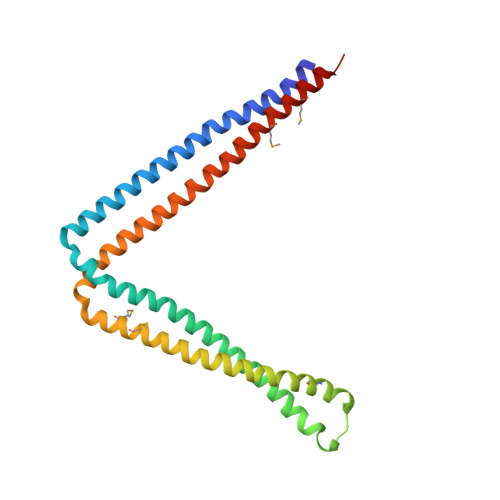
6IXF

Crystal structure of SeMet apo SH3BP5 (P41)


Experimental Data Snapshot

  • Method: X-RAY DIFFRACTION
  • Resolution: 3.60 Å
  • R-Value Free: 0.261 
  • R-Value Work: 0.234 
  • R-Value Observed: 0.235 

Starting Model: experimental
View more details

wwPDB Validation   3D Report Full Report


This is version 1.3 of the entry. See complete history


Literature

Structural basis of guanine nucleotide exchange for Rab11 by SH3BP5.

Goto-Ito, S.Morooka, N.Yamagata, A.Sato, Y.Sato, K.Fukai, S.

(2019) Life Sci Alliance 2

  • DOI: https://doi.org/10.26508/lsa.201900297
  • Primary Citation of Related Structures:  
    6IXE, 6IXF, 6IXG, 6IXV

  • PubMed Abstract: 

    The Rab GTPase family is a major regulator of membrane traffic in eukaryotic cells. The Rab11 subfamily plays important roles in specific trafficking events such as exocytosis, endosomal recycling, and cytokinesis. SH3BP5 and SH3BP5-like (SH3BP5L) proteins have recently been found to serve as guanine nucleotide exchange factors (GEF) for Rab11. Here, we report the crystal structures of the SH3BP5 GEF domain alone and its complex with Rab11a. SH3BP5 exhibits a V-shaped structure comprising two coiled coils. The coiled coil composed of α1, and α4 is solely responsible for the Rab11a binding and GEF activity. SH3BP5 pulls out and deforms switch I of Rab11a so as to facilitate the GDP release from Rab11a. SH3BP5 interacts with the N-terminal region, switch I, interswitch, and switch II of Rab11a. SH3BP5 and SH3BP5L localize to Rab11-positive recycling endosomes and show GEF activity for all of the Rab11 family but not for Rab14. Fluorescence-based GEF assays combined with site-directed mutagenesis reveal the essential interactions between SH3BP5 and Rab11 family proteins for the GEF reaction on recycling endosomes.


  • Organizational Affiliation

    Institute for Quantitative Biosciences, The University of Tokyo, Tokyo, Japan.


Macromolecules
Find similar proteins by:  (by identity cutoff)  |  3D Structure
Entity ID: 1
MoleculeChains Sequence LengthOrganismDetailsImage
SH3 domain-binding protein 5
A, B
228Homo sapiensMutation(s): 4 
Gene Names: SH3BP5SAB
UniProt & NIH Common Fund Data Resources
Find proteins for O60239 (Homo sapiens)
Explore O60239 
Go to UniProtKB:  O60239
PHAROS:  O60239
GTEx:  ENSG00000131370 
Entity Groups  
Sequence Clusters30% Identity50% Identity70% Identity90% Identity95% Identity100% Identity
UniProt GroupO60239
Sequence Annotations
Expand
  • Reference Sequence
Small Molecules
Modified Residues  1 Unique
IDChains TypeFormula2D DiagramParent
MSE
Query on MSE
A, B
L-PEPTIDE LINKINGC5 H11 N O2 SeMET
Experimental Data & Validation

Experimental Data

  • Method: X-RAY DIFFRACTION
  • Resolution: 3.60 Å
  • R-Value Free: 0.261 
  • R-Value Work: 0.234 
  • R-Value Observed: 0.235 
  • Space Group: P 41
Unit Cell:
Length ( Å )Angle ( ˚ )
a = 78.212α = 90
b = 78.212β = 90
c = 92.964γ = 90
Software Package:
Software NamePurpose
PHENIXrefinement
HKL-2000data reduction
HKL-2000data scaling
PHASERphasing

Structure Validation

View Full Validation Report


Entry History & Funding Information

Deposition Data


Funding OrganizationLocationGrant Number
Japan Society for the Promotion of ScienceJapan17K15072

Revision History  (Full details and data files)

  • Version 1.0: 2019-03-20
    Type: Initial release
  • Version 1.1: 2019-03-27
    Changes: Data collection, Database references
  • Version 1.2: 2023-11-22
    Changes: Data collection, Database references, Refinement description
  • Version 1.3: 2024-11-13
    Changes: Structure summary