6CQ9

K2P2.1(TREK-1):ML402 complex


Experimental Data Snapshot

  • Method: X-RAY DIFFRACTION
  • Resolution: 2.80 Å
  • R-Value Free: 0.309 
  • R-Value Work: 0.265 
  • R-Value Observed: 0.267 

Starting Model: experimental
View more details

wwPDB Validation   3D Report Full Report


Ligand Structure Quality Assessment 

Created with Raphaël 2.3.0Worse 01 BetterLigand structure goodness of fit to experimental dataBest fitted 16CClick on this verticalbar to view detailsBest fitted Q5FClick on this verticalbar to view detailsBest fitted R16Click on this verticalbar to view details

This is version 1.5 of the entry. See complete history


Literature

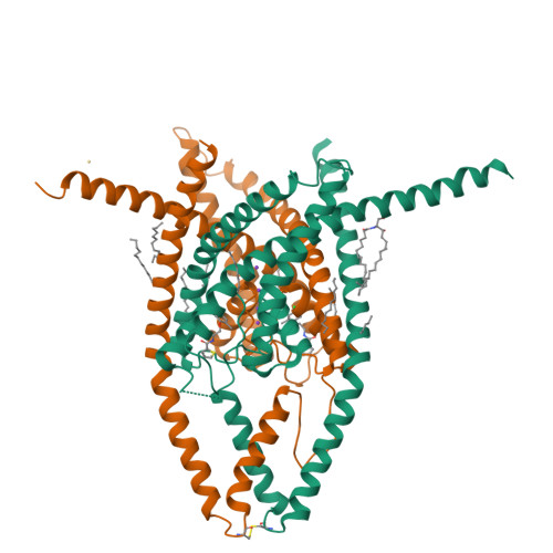
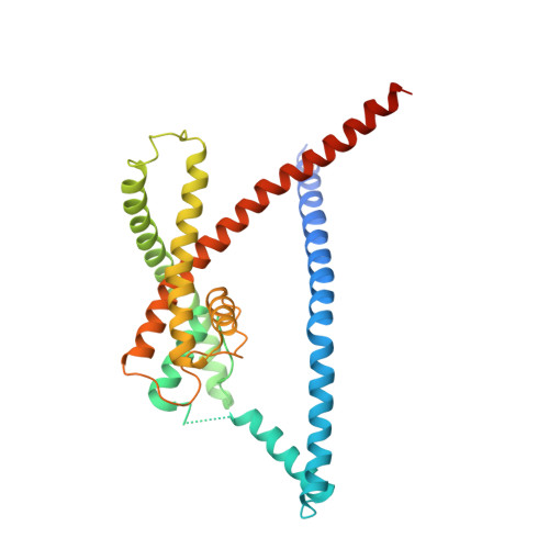
K2P2.1 (TREK-1)-activator complexes reveal a cryptic selectivity filter binding site.

Lolicato, M.Arrigoni, C.Mori, T.Sekioka, Y.Bryant, C.Clark, K.A.Minor, D.L.

(2017) Nature 547: 364-368

  • DOI: https://doi.org/10.1038/nature22988
  • Primary Citation of Related Structures:  
    6CQ6, 6CQ8, 6CQ9

  • PubMed Abstract: 

    Polymodal thermo- and mechanosensitive two-pore domain potassium (K 2P ) channels of the TREK subfamily generate 'leak' currents that regulate neuronal excitability, respond to lipids, temperature and mechanical stretch, and influence pain, temperature perception and anaesthetic responses. These dimeric voltage-gated ion channel (VGIC) superfamily members have a unique topology comprising two pore-forming regions per subunit. In contrast to other potassium channels, K 2P channels use a selectivity filter 'C-type' gate as the principal gating site. Despite recent advances, poor pharmacological profiles of K 2P channels limit mechanistic and biological studies. Here we describe a class of small-molecule TREK activators that directly stimulate the C-type gate by acting as molecular wedges that restrict interdomain interface movement behind the selectivity filter. Structures of K 2P 2.1 (also known as TREK-1) alone and with two selective K 2P 2.1 (TREK-1) and K 2P 10.1 (TREK-2) activators-an N-aryl-sulfonamide, ML335, and a thiophene-carboxamide, ML402-define a cryptic binding pocket unlike other ion channel small-molecule binding sites and, together with functional studies, identify a cation-π interaction that controls selectivity. Together, our data reveal a druggable K 2P site that stabilizes the C-type gate 'leak mode' and provide direct evidence for K 2P selectivity filter gating.


  • Organizational Affiliation

    Cardiovascular Research Institute, University of California, San Francisco, California 941158-9001, USA.


Macromolecules
Find similar proteins by:  (by identity cutoff)  |  3D Structure
Entity ID: 1
MoleculeChains Sequence LengthOrganismDetailsImage
Potassium channel subfamily K member 2
A, B
312Mus musculusMutation(s): 15 
Gene Names: Kcnk2
Membrane Entity: Yes 
UniProt & NIH Common Fund Data Resources
Find proteins for P97438 (Mus musculus)
Explore P97438 
Go to UniProtKB:  P97438
IMPC:  MGI:109366
Entity Groups  
Sequence Clusters30% Identity50% Identity70% Identity90% Identity95% Identity100% Identity
UniProt GroupP97438
Sequence Annotations
Expand
  • Reference Sequence
Small Molecules
Ligands 5 Unique
IDChains Name / Formula / InChI Key2D Diagram3D Interactions
16C
Query on 16C

Download Ideal Coordinates CCD File 
H [auth A]N-((E,2S,3R)-1,3-DIHYDROXYOCTADEC-4-EN-2-YL)PALMITAMIDE
C34 H67 N O3
YDNKGFDKKRUKPY-TURZORIXSA-N
Q5F
Query on Q5F

Download Ideal Coordinates CCD File 
F [auth A],
N [auth B]
N-[2-(4-chloro-2-methylphenoxy)ethyl]thiophene-2-carboxamide
C14 H14 Cl N O2 S
RULQUKFOBAPKKR-UHFFFAOYSA-N
R16
Query on R16

Download Ideal Coordinates CCD File 
G [auth A]
I [auth A]
J [auth A]
O [auth B]
P [auth B]
HEXADECANE
C16 H34
DCAYPVUWAIABOU-UHFFFAOYSA-N
CD
Query on CD

Download Ideal Coordinates CCD File 
M [auth B]CADMIUM ION
Cd
WLZRMCYVCSSEQC-UHFFFAOYSA-N
K
Query on K

Download Ideal Coordinates CCD File 
C [auth A],
D [auth A],
E [auth A],
K [auth B],
L [auth B]
POTASSIUM ION
K
NPYPAHLBTDXSSS-UHFFFAOYSA-N
Experimental Data & Validation

Experimental Data

  • Method: X-RAY DIFFRACTION
  • Resolution: 2.80 Å
  • R-Value Free: 0.309 
  • R-Value Work: 0.265 
  • R-Value Observed: 0.267 
  • Space Group: P 21 21 21
Unit Cell:
Length ( Å )Angle ( ˚ )
a = 67.091α = 90
b = 119.565β = 90
c = 127.215γ = 90
Software Package:
Software NamePurpose
PHENIXrefinement
Cootmodel building
Aimlessdata scaling
PHASERphasing
XDSdata processing
XDSdata reduction

Structure Validation

View Full Validation Report


Ligand Structure Quality Assessment 

Created with Raphaël 2.3.0Worse 01 BetterLigand structure goodness of fit to experimental dataBest fitted 16CClick on this verticalbar to view detailsBest fitted Q5FClick on this verticalbar to view detailsBest fitted R16Click on this verticalbar to view details

Entry History & Funding Information

Deposition Data


Funding OrganizationLocationGrant Number
National Institutes of Health/National Institute of Mental Health (NIH/NIMH)United States--
American Heart AssociationUnited States--

Revision History  (Full details and data files)

  • Version 1.0: 2018-04-11
    Type: Initial release
  • Version 1.1: 2019-02-20
    Changes: Author supporting evidence, Data collection
  • Version 1.2: 2019-11-06
    Changes: Author supporting evidence, Data collection
  • Version 1.3: 2019-11-27
    Changes: Author supporting evidence
  • Version 1.4: 2023-10-04
    Changes: Data collection, Database references, Derived calculations, Refinement description, Structure summary
  • Version 1.5: 2024-11-13
    Changes: Structure summary