5OQU

The crystal structure of CK2alpha in complex with compound 5


Experimental Data Snapshot

  • Method: X-RAY DIFFRACTION
  • Resolution: 2.32 Å
  • R-Value Free: 0.244 
  • R-Value Work: 0.220 
  • R-Value Observed: 0.221 

Starting Model: experimental
View more details

wwPDB Validation   3D Report Full Report


Ligand Structure Quality Assessment 

Created with Raphaël 2.3.0Worse 01 BetterLigand structure goodness of fit to experimental dataBest fitted IHPClick on this verticalbar to view detailsBest fitted ADPClick on this verticalbar to view detailsBest fitted A4BClick on this verticalbar to view details

This is version 2.1 of the entry. See complete history


Literature

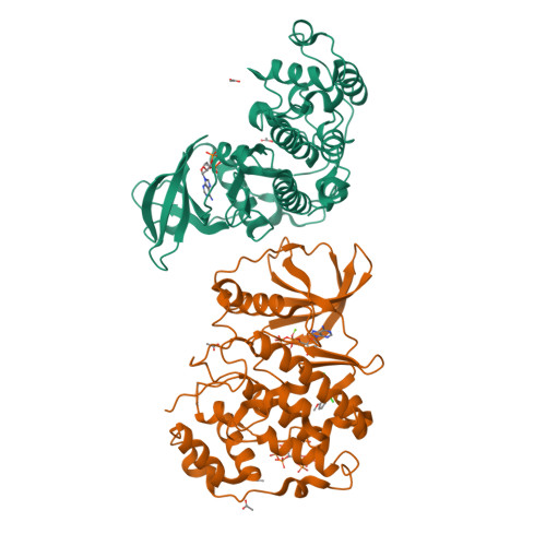
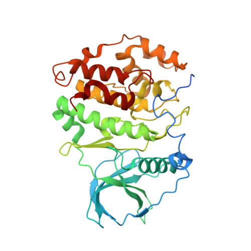
Second-generation CK2 alpha inhibitors targeting the alpha D pocket.

Iegre, J.Brear, P.De Fusco, C.Yoshida, M.Mitchell, S.L.Rossmann, M.Carro, L.Sore, H.F.Hyvonen, M.Spring, D.R.

(2018) Chem Sci 9: 3041-3049

  • DOI: https://doi.org/10.1039/c7sc05122k
  • Primary Citation of Related Structures:  
    5OQU, 5ORH, 5ORJ, 5ORK, 5OS7, 5OS8, 5OSL, 5OSP, 5OSR, 5OSU, 5OSZ, 5OT5, 5OT6, 5OTD, 5OTH, 5OTI, 5OTL, 5OTO, 5OTP, 5OTQ, 5OTR, 5OTS, 5OTY, 5OTZ, 5OUE, 5OUL, 5OUM, 5OUU, 5OYF, 6EHK, 6EHU, 6EII

  • PubMed Abstract: 

    CK2 is a critical cell cycle regulator that also promotes various anti-apoptotic mechanisms. Development of ATP-non-competitive inhibitors of CK2 is a very attractive strategy considering that the ATP binding site is highly conserved among other kinases. We have previously utilised a pocket outside the active site to develop a novel CK2 inhibitor, CAM4066 . Whilst CAM4066 bound to this new pocket it was also interacting with the ATP site: herein, we describe an example of a CK2α inhibitor that binds completely outside the active site. This second generation αD-site binding inhibitor, compound CAM4712 (IC 50 = 7 μM, GI 50 = 10.0 ± 3.6 μM), has numerous advantages over the previously reported CAM4066 , including a reduction in the number of rotatable bonds, the absence of amide groups susceptible to the action of proteases and improved cellular permeability. Unlike with CAM4066 , there was no need to facilitate cellular uptake by making a prodrug. Moreover, CAM4712 displayed no drop off between its ability to inhibit the kinase in vitro (IC 50 ) and the ability to inhibit cell proliferation (GI 50 ).


  • Organizational Affiliation

    Department of Chemistry , University of Cambridge , CB2 1EW , Cambridge , UK . Email: spring@ch.cam.ac.uk.


Macromolecules
Find similar proteins by:  (by identity cutoff)  |  3D Structure
Entity ID: 1
MoleculeChains Sequence LengthOrganismDetailsImage
Casein kinase II subunit alphaA [auth B],
B [auth A]
352Homo sapiensMutation(s): 1 
Gene Names: CSNK2A1CK2A1
EC: 2.7.11.1
UniProt & NIH Common Fund Data Resources
Find proteins for P68400 (Homo sapiens)
Explore P68400 
Go to UniProtKB:  P68400
PHAROS:  P68400
GTEx:  ENSG00000101266 
Entity Groups  
Sequence Clusters30% Identity50% Identity70% Identity90% Identity95% Identity100% Identity
UniProt GroupP68400
Sequence Annotations
Expand
  • Reference Sequence
Small Molecules
Ligands 6 Unique
IDChains Name / Formula / InChI Key2D Diagram3D Interactions
IHP
Query on IHP

Download Ideal Coordinates CCD File 
M [auth A]INOSITOL HEXAKISPHOSPHATE
C6 H18 O24 P6
IMQLKJBTEOYOSI-GPIVLXJGSA-N
ADP
Query on ADP

Download Ideal Coordinates CCD File 
C [auth B],
F [auth A]
ADENOSINE-5'-DIPHOSPHATE
C10 H15 N5 O10 P2
XTWYTFMLZFPYCI-KQYNXXCUSA-N
A4B
Query on A4B

Download Ideal Coordinates CCD File 
G [auth A][3-chloranyl-4-(2-methoxyphenyl)phenyl]methanamine
C14 H14 Cl N O
LRYGKGSWWOPVKM-UHFFFAOYSA-N
ACT
Query on ACT

Download Ideal Coordinates CCD File 
D [auth B],
E [auth B],
J [auth A],
K [auth A],
L [auth A]
ACETATE ION
C2 H3 O2
QTBSBXVTEAMEQO-UHFFFAOYSA-M
CL
Query on CL

Download Ideal Coordinates CCD File 
N [auth A]CHLORIDE ION
Cl
VEXZGXHMUGYJMC-UHFFFAOYSA-M
MG
Query on MG

Download Ideal Coordinates CCD File 
H [auth A],
I [auth A]
MAGNESIUM ION
Mg
JLVVSXFLKOJNIY-UHFFFAOYSA-N
Binding Affinity Annotations 
IDSourceBinding Affinity
A4B BindingDB:  5OQU IC50: 4.93e+5 (nM) from 1 assay(s)
Experimental Data & Validation

Experimental Data

  • Method: X-RAY DIFFRACTION
  • Resolution: 2.32 Å
  • R-Value Free: 0.244 
  • R-Value Work: 0.220 
  • R-Value Observed: 0.221 
  • Space Group: C 2 2 21
Unit Cell:
Length ( Å )Angle ( ˚ )
a = 64.668α = 90
b = 67.922β = 90
c = 332.804γ = 90
Software Package:
Software NamePurpose
BUSTERrefinement
Aimlessdata scaling
PDB_EXTRACTdata extraction
PHASERphasing

Structure Validation

View Full Validation Report


Ligand Structure Quality Assessment 

Created with Raphaël 2.3.0Worse 01 BetterLigand structure goodness of fit to experimental dataBest fitted IHPClick on this verticalbar to view detailsBest fitted ADPClick on this verticalbar to view detailsBest fitted A4BClick on this verticalbar to view details

Entry History & Funding Information

Deposition Data


Funding OrganizationLocationGrant Number
Wellcome TrustUnited Kingdom090340/Z/09/Z
Wellcome TrustUnited Kingdom107714/Z/15/Z

Revision History  (Full details and data files)

  • Version 1.0: 2018-02-28
    Type: Initial release
  • Version 1.1: 2018-05-16
    Changes: Data collection, Database references
  • Version 2.0: 2020-10-07
    Changes: Advisory, Atomic model, Data collection, Derived calculations, Non-polymer description, Refinement description, Structure summary
  • Version 2.1: 2024-01-17
    Changes: Data collection, Database references, Refinement description