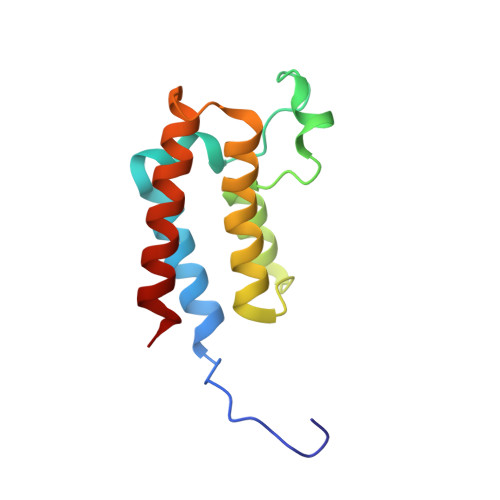
5MGF

Crystal Structure of BAZ2B bromodomain in complex with 4-propionyl-pyrrole derivative 2


Experimental Data Snapshot

  • Method: X-RAY DIFFRACTION
  • Resolution: 1.90 Å
  • R-Value Free: 0.201 
  • R-Value Work: 0.177 
  • R-Value Observed: 0.178 

Starting Model: experimental
View more details

wwPDB Validation   3D Report Full Report


Ligand Structure Quality Assessment 


This is version 1.2 of the entry. See complete history


Literature

Discovery of BAZ2A bromodomain ligands.

Spiliotopoulos, D.Wamhoff, E.C.Lolli, G.Rademacher, C.Caflisch, A.

(2017) Eur J Med Chem 139: 564-572

  • DOI: https://doi.org/10.1016/j.ejmech.2017.08.028
  • Primary Citation of Related Structures:  
    5MGE, 5MGF, 5MGG, 5MGJ, 5MGK, 5MGL, 5MGM

  • PubMed Abstract: 

    The bromodomain adjacent to zinc finger domain protein 2A (BAZ2A) is implicated in aggressive prostate cancer. The BAZ2A bromodomain is a challenging target because of the shallow pocket of its natural ligand, the acetylated side chain of lysine. Here, we report the successful screening of a library of nearly 1500 small molecules by high-throughput docking and force field-based binding-energy evaluation. For seven of the 20 molecules selected in silico, evidence of binding to the BAZ2A bromodomain is provided by ligand-observed NMR spectroscopy. Two of these compounds show a favorable ligand efficiency of 0.42 kcal/mol per non-hydrogen atom in a competition-binding assay. The crystal structures of the BAZ2A bromodomain in complex with four fragment hits validate the predicted binding modes. The binding modes of compounds 1 and 3 are compatible with ligand growing for optimization of affinity for BAZ2A and selectivity against the close homologue BAZ2B.


  • Organizational Affiliation

    Department of Biochemistry, University of Zürich, Winterthurerstrasse 190, CH-8057 Zürich, Switzerland.


Macromolecules
Find similar proteins by:  (by identity cutoff)  |  3D Structure
Entity ID: 1
MoleculeChains Sequence LengthOrganismDetailsImage
Bromodomain adjacent to zinc finger domain protein 2B116Homo sapiensMutation(s): 0 
Gene Names: BAZ2BKIAA1476
UniProt & NIH Common Fund Data Resources
Find proteins for Q9UIF8 (Homo sapiens)
Explore Q9UIF8 
Go to UniProtKB:  Q9UIF8
PHAROS:  Q9UIF8
GTEx:  ENSG00000123636 
Entity Groups  
Sequence Clusters30% Identity50% Identity70% Identity90% Identity95% Identity100% Identity
UniProt GroupQ9UIF8
Sequence Annotations
Expand
  • Reference Sequence
Small Molecules
Ligands 1 Unique
IDChains Name / Formula / InChI Key2D Diagram3D Interactions
7MW
Query on 7MW

Download Ideal Coordinates CCD File 
B [auth A]methyl 4-propanoyl-1~{H}-pyrrole-2-carboxylate
C9 H11 N O3
GMDIDIYLWBUNEW-UHFFFAOYSA-N
Binding Affinity Annotations 
IDSourceBinding Affinity
7MW BindingDB:  5MGF Kd: 5.40e+4 (nM) from 1 assay(s)
Experimental Data & Validation

Experimental Data

  • Method: X-RAY DIFFRACTION
  • Resolution: 1.90 Å
  • R-Value Free: 0.201 
  • R-Value Work: 0.177 
  • R-Value Observed: 0.178 
  • Space Group: C 2 2 21
Unit Cell:
Length ( Å )Angle ( ˚ )
a = 81.118α = 90
b = 96.103β = 90
c = 57.48γ = 90
Software Package:
Software NamePurpose
Aimlessdata scaling
XDSdata reduction
PHENIXrefinement
PDB_EXTRACTdata extraction
XSCALEdata scaling
PHASERphasing

Structure Validation

View Full Validation Report


Ligand Structure Quality Assessment 


Entry History & Funding Information

Deposition Data


Funding OrganizationLocationGrant Number
Swiss National Science FoundationSwitzerland31003A_169007

Revision History  (Full details and data files)

  • Version 1.0: 2017-04-12
    Type: Initial release
  • Version 1.1: 2017-09-06
    Changes: Data collection, Database references, Refinement description
  • Version 1.2: 2024-01-17
    Changes: Data collection, Database references, Refinement description