5IWE

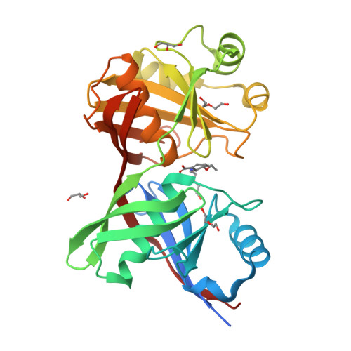
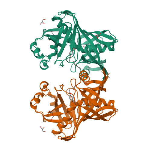
E45Q mutant of phenazine biosynthesis protein PhzF in complex with (5R,6R)-6-azaniumyl-5-ethoxycyclohexa-1,3-diene-1-carboxylate


Experimental Data Snapshot

  • Method: X-RAY DIFFRACTION
  • Resolution: 1.71 Å
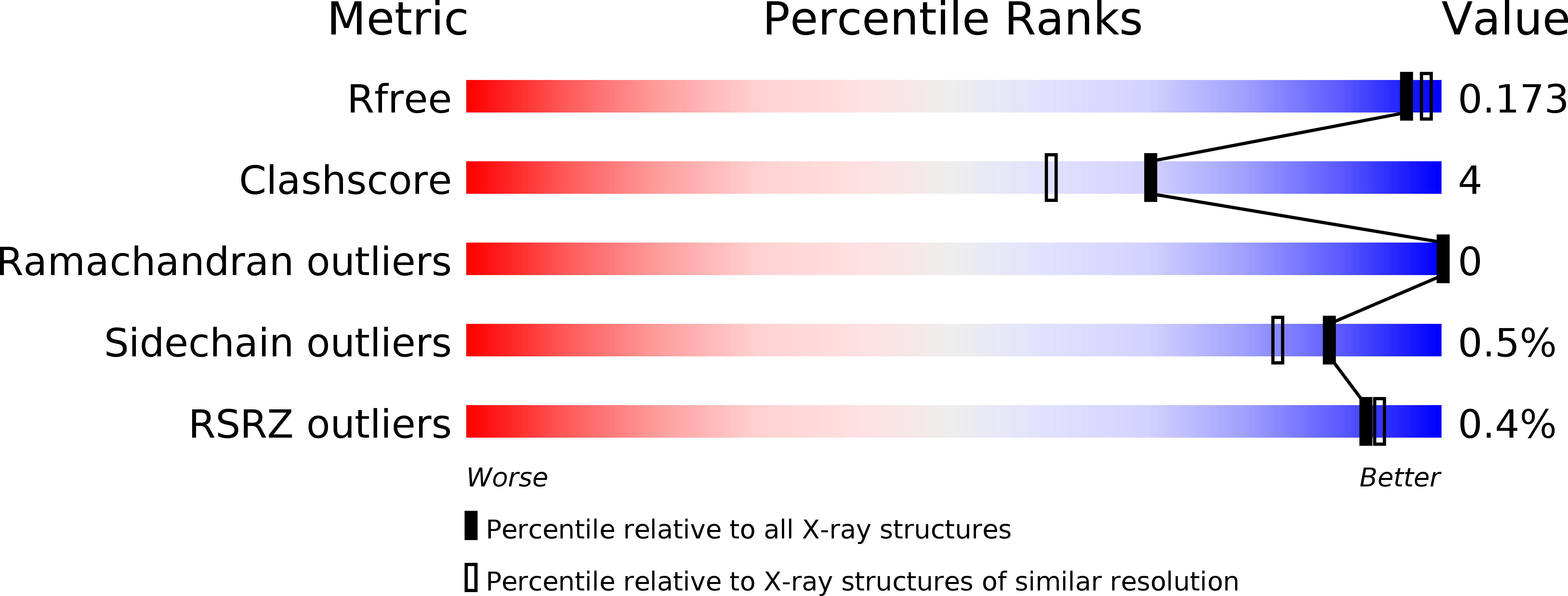
  • R-Value Free: 0.173 
  • R-Value Work: 0.149 
  • R-Value Observed: 0.150 

Starting Model: experimental
View more details

wwPDB Validation   3D Report Full Report


Ligand Structure Quality Assessment 


This is version 1.2 of the entry. See complete history


Literature

Mechanisms and Specificity of Phenazine Biosynthesis Protein PhzF.

Diederich, C.Leypold, M.Culka, M.Weber, H.Breinbauer, R.Ullmann, G.M.Blankenfeldt, W.

(2017) Sci Rep 7: 6272-6272

  • DOI: https://doi.org/10.1038/s41598-017-06278-w
  • Primary Citation of Related Structures:  
    5IWE

  • PubMed Abstract: 

    Phenazines are bacterial virulence and survival factors with important roles in infectious disease. PhzF catalyzes a key reaction in their biosynthesis by isomerizing (2 S,3 S)-2,3-dihydro-3-hydroxy anthranilate (DHHA) in two steps, a [1,5]-hydrogen shift followed by tautomerization to an aminoketone. While the [1,5]-hydrogen shift requires the conserved glutamate E45, suggesting acid/base catalysis, it also shows hallmarks of a sigmatropic rearrangement, namely the suprafacial migration of a non-acidic proton. To discriminate these mechanistic alternatives, we employed enzyme kinetic measurements and computational methods. Quantum mechanics/molecular mechanics (QM/MM) calculations revealed that the activation barrier of a proton shuttle mechanism involving E45 is significantly lower than that of a sigmatropic [1,5]-hydrogen shift. QM/MM also predicted a large kinetic isotope effect, which was indeed observed with deuterated substrate. For the tautomerization, QM/MM calculations suggested involvement of E45 and an active site water molecule, explaining the observed stereochemistry. Because these findings imply that PhzF can act only on a limited substrate spectrum, we also investigated the turnover of DHHA derivatives, of which only O-methyl and O-ethyl DHHA were converted. Together, these data reveal how PhzF orchestrates a water-free with a water-dependent step. Its unique mechanism, specificity and essential role in phenazine biosynthesis may offer opportunities for inhibitor development.


  • Organizational Affiliation

    Structure and Function of Proteins, Helmholtz Centre for Infection Research, Inhoffenstr. 7, 38124, Braunschweig, Germany.


Macromolecules
Find similar proteins by:  (by identity cutoff)  |  3D Structure
Entity ID: 1
MoleculeChains Sequence LengthOrganismDetailsImage
Trans-2,3-dihydro-3-hydroxyanthranilate isomerase298Pseudomonas fluorescensMutation(s): 1 
Gene Names: phzF
EC: 5.3.3.17
UniProt
Find proteins for Q51792 (Pseudomonas fluorescens)
Explore Q51792 
Go to UniProtKB:  Q51792
Entity Groups  
Sequence Clusters30% Identity50% Identity70% Identity90% Identity95% Identity100% Identity
UniProt GroupQ51792
Sequence Annotations
Expand
  • Reference Sequence
Small Molecules
Ligands 4 Unique
IDChains Name / Formula / InChI Key2D Diagram3D Interactions
W81
Query on W81

Download Ideal Coordinates CCD File 
B [auth A](5R,6R)-6-azaniumyl-5-ethoxycyclohexa-1,3-diene-1-carboxylate
C9 H13 N O3
UOFNVIJXMFQHDJ-HTQZYQBOSA-N
PEG
Query on PEG

Download Ideal Coordinates CCD File 
C [auth A],
D [auth A]
DI(HYDROXYETHYL)ETHER
C4 H10 O3
MTHSVFCYNBDYFN-UHFFFAOYSA-N
GOL
Query on GOL

Download Ideal Coordinates CCD File 
G [auth A],
H [auth A]
GLYCEROL
C3 H8 O3
PEDCQBHIVMGVHV-UHFFFAOYSA-N
EDO
Query on EDO

Download Ideal Coordinates CCD File 
E [auth A],
F [auth A]
1,2-ETHANEDIOL
C2 H6 O2
LYCAIKOWRPUZTN-UHFFFAOYSA-N
Experimental Data & Validation

Experimental Data

  • Method: X-RAY DIFFRACTION
  • Resolution: 1.71 Å
  • R-Value Free: 0.173 
  • R-Value Work: 0.149 
  • R-Value Observed: 0.150 
  • Space Group: P 32 2 1
Unit Cell:
Length ( Å )Angle ( ˚ )
a = 56.032α = 90
b = 56.032β = 90
c = 155.742γ = 120
Software Package:
Software NamePurpose
PHENIXrefinement
XDSdata reduction
Aimlessdata scaling
PHASERphasing

Structure Validation

View Full Validation Report


Ligand Structure Quality Assessment 


Entry History & Funding Information

Deposition Data


Funding OrganizationLocationGrant Number
ERA-Chemistry networkGermanyBL587-3
ERA-Chemistry networkAustriaI-668

Revision History  (Full details and data files)

  • Version 1.0: 2017-03-29
    Type: Initial release
  • Version 1.1: 2017-08-09
    Changes: Database references
  • Version 1.2: 2024-01-10
    Changes: Data collection, Database references, Refinement description