5HTB

Crystal structure of haspin (GSG2) in complex with bisubstrate inhibitor ARC-3353


Experimental Data Snapshot

  • Method: X-RAY DIFFRACTION
  • Resolution: 1.70 Å
  • R-Value Free: 
    0.181 (Depositor), 0.190 (DCC) 
  • R-Value Work: 
    0.162 (Depositor), 0.173 (DCC) 
  • R-Value Observed: 
    0.163 (Depositor) 

wwPDB Validation   3D Report Full Report


Ligand Structure Quality Assessment 

Created with Raphaël 2.3.0Worse 01 BetterLigand structure goodness of fit to experimental dataBest fitted 6L5Click on this verticalbar to view details

This is version 2.0 of the entry. See complete history


Literature

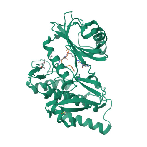
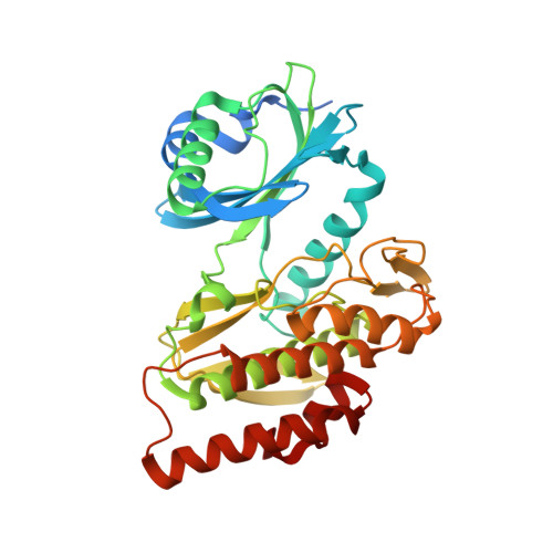
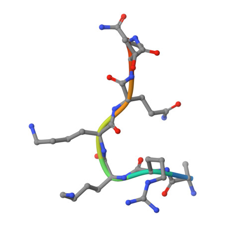
Co-crystal structures of the protein kinase haspin with bisubstrate inhibitors.

Lavogina, D.Kestav, K.Chaikuad, A.Heroven, C.Knapp, S.Uri, A.

(2016) Acta Crystallogr F Struct Biol Commun 72: 339-345

  • DOI: https://doi.org/10.1107/S2053230X16004611
  • Primary Citation of Related Structures:  
    5HTB, 5HTC

  • PubMed Abstract: 

    Haspin is a mitotic protein kinase that is responsible for the phosphorylation of Thr3 of histone H3, thereby creating a recognition motif for docking of the chromosomal passenger complex that is crucial for the progression of cell division. Here, two high-resolution models of haspin with previously reported inhibitors consisting of an ATP analogue and a histone H3(1-7) peptide analogue are presented. The structures of the complexes confirm the bisubstrate character of the inhibitors by revealing the signature binding modes of the moieties targeting the ATP-binding site and the protein substrate-binding site of the kinase. This is the first structural model of a bisubstrate inhibitor targeting haspin. The presented structural data represent a model for the future development of more specific haspin inhibitors.


  • Organizational Affiliation

    Institute of Chemistry, University of Tartu, Ravila 14A, 50411 Tartu, Estonia.


Macromolecules
Find similar proteins by:  (by identity cutoff)  |  3D Structure
Entity ID: 1
MoleculeChains Sequence LengthOrganismDetailsImage
Serine/threonine-protein kinase haspin357Homo sapiensMutation(s): 0 
Gene Names: GSG2
EC: 2.7.11.1
UniProt & NIH Common Fund Data Resources
Find proteins for Q8TF76 (Homo sapiens)
Explore Q8TF76 
Go to UniProtKB:  Q8TF76
PHAROS:  Q8TF76
GTEx:  ENSG00000177602 
Entity Groups  
Sequence Clusters30% Identity50% Identity70% Identity90% Identity95% Identity100% Identity
UniProt GroupQ8TF76
Sequence Annotations
Expand
  • Reference Sequence

Find similar proteins by:  Sequence   |   3D Structure  

Entity ID: 2
MoleculeChains Sequence LengthOrganismDetailsImage
(2R,3S,4R,5R,6R)-6-((1R,2R,3S,4R,6S)-4,6-DIAMINO-2,3-DIHYDROXYCYCLOHEXYLOXY)-5-AMINO-2-(AMINOMETHYL)-TETRAHYDRO-2H-PYRAN-3,4-DIOLB [auth C]8synthetic constructMutation(s): 0 
Sequence Annotations
Expand
  • Reference Sequence
Small Molecules
Ligands 5 Unique
IDChains Name / Formula / InChI Key2D Diagram3D Interactions
6L5
Query on 6L5

Download Ideal Coordinates CCD File 
F [auth C](3R)-4-amino-3-{[6-({[(2S,3S,4R,5R)-5-(6-amino-9H-purin-9-yl)-3,4-dihydroxytetrahydrofuran-2-yl]carbonyl}amino)hexanoyl]amino}-4-oxobutanoic acid (non-preferred name)
C20 H28 N8 O8
WWJTVYJOLVMSCT-BRLJYLSVSA-N
MPD
Query on MPD

Download Ideal Coordinates CCD File 
G [auth C](4S)-2-METHYL-2,4-PENTANEDIOL
C6 H14 O2
SVTBMSDMJJWYQN-YFKPBYRVSA-N
MRD
Query on MRD

Download Ideal Coordinates CCD File 
C [auth A](4R)-2-METHYLPENTANE-2,4-DIOL
C6 H14 O2
SVTBMSDMJJWYQN-RXMQYKEDSA-N
DMS
Query on DMS

Download Ideal Coordinates CCD File 
E [auth A]DIMETHYL SULFOXIDE
C2 H6 O S
IAZDPXIOMUYVGZ-UHFFFAOYSA-N
NA
Query on NA

Download Ideal Coordinates CCD File 
D [auth A]SODIUM ION
Na
FKNQFGJONOIPTF-UHFFFAOYSA-N
Experimental Data & Validation

Experimental Data

  • Method: X-RAY DIFFRACTION
  • Resolution: 1.70 Å
  • R-Value Free:  0.181 (Depositor), 0.190 (DCC) 
  • R-Value Work:  0.162 (Depositor), 0.173 (DCC) 
  • R-Value Observed: 0.163 (Depositor) 
Space Group: P 21 21 21
Unit Cell:
Length ( Å )Angle ( ˚ )
a = 77.79α = 90
b = 78.89β = 90
c = 81.75γ = 90
Software Package:
Software NamePurpose
REFMACrefinement
iMOSFLMdata reduction
SCALAdata scaling
PHASERphasing

Structure Validation

View Full Validation Report


Ligand Structure Quality Assessment 

Created with Raphaël 2.3.0Worse 01 BetterLigand structure goodness of fit to experimental dataBest fitted 6L5Click on this verticalbar to view details

Entry History 

Revision History  (Full details and data files)

  • Version 1.0: 2016-05-11
    Type: Initial release
  • Version 2.0: 2024-12-04
    Changes: Advisory, Atomic model, Data collection, Database references, Derived calculations, Non-polymer description, Polymer sequence, Source and taxonomy, Structure summary