5B49

Crystal structure of LpxH with manganese from Pseudomonas aeruginosa


Experimental Data Snapshot

  • Method: X-RAY DIFFRACTION
  • Resolution: 1.65 Å
  • R-Value Free: 
    0.177 (Depositor), 0.190 (DCC) 
  • R-Value Work: 
    0.152 (Depositor), 0.160 (DCC) 
  • R-Value Observed: 
    0.153 (Depositor) 

wwPDB Validation   3D Report Full Report


Ligand Structure Quality Assessment 

Created with Raphaël 2.3.0Worse 01 BetterLigand structure goodness of fit to experimental dataBest fitted LP5Click on this verticalbar to view details

This is version 1.2 of the entry. See complete history


Literature

Crystal structures of the UDP-diacylglucosamine pyrophosphohydrase LpxH from Pseudomonas aeruginosa

Okada, C.Wakabayashi, H.Kobayashi, M.Shinoda, A.Tanaka, I.Yao, M.

(2016) Sci Rep 6: 32822-32822

  • DOI: https://doi.org/10.1038/srep32822
  • Primary Citation of Related Structures:  
    5B49, 5B4A, 5B4B, 5B4C, 5B4D

  • PubMed Abstract: 

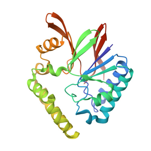
    Lipid A (also known as endotoxin) is the hydrophobic portion of lipopolysaccharides. It is an essential membrane component required for the viability of gram-negative bacteria. The enzymes involved in its biosynthesis are attractive targets for the development of novel antibiotics. LpxH catalyzes the fourth step of the lipid A biosynthesis pathway and cleaves the pyrophosphate bond of UDP-2,3-diacylglucosamine to yield 2,3-diacylglucosamine 1-phosphate (lipid X) and UMP. Here we present the structures of LpxH from Pseudomonas aeruginosa (PaLpxH). PaLpxH consists of two domains: a catalytic domain that is homologous to the metallophosphoesterases and a helical insertion domain. Lipid X was captured in the crevice between these two domains, with its phosphate group facing the dinuclear metal (Mn(2+)) center and two acyl chains buried in the hydrophobic cavity. The structures reveal that a large conformational change occurs at the lipid X binding site surface upon the binding/release of the product molecule. Based on these observations, we propose a novel model for lipid X embedding, which involves the scissor-like movement of helix α6, resulting in the release of lipid X into the lipid bilayer.


  • Organizational Affiliation

    Faculty of Advanced Life Science, Hokkaido University, Sapporo 060-0810, Japan.


Macromolecules
Find similar proteins by:  (by identity cutoff)  |  3D Structure
Entity ID: 1
MoleculeChains Sequence LengthOrganismDetailsImage
UDP-2,3-diacylglucosamine hydrolase
A, B
248Pseudomonas aeruginosa PAO1Mutation(s): 0 
Gene Names: lpxHPA1792
EC: 3.6.1.54
Membrane Entity: Yes 
UniProt
Find proteins for Q9I2V0 (Pseudomonas aeruginosa (strain ATCC 15692 / DSM 22644 / CIP 104116 / JCM 14847 / LMG 12228 / 1C / PRS 101 / PAO1))
Explore Q9I2V0 
Go to UniProtKB:  Q9I2V0
Entity Groups  
Sequence Clusters30% Identity50% Identity70% Identity90% Identity95% Identity100% Identity
UniProt GroupQ9I2V0
Sequence Annotations
Expand
  • Reference Sequence
Small Molecules
Ligands 3 Unique
IDChains Name / Formula / InChI Key2D Diagram3D Interactions
LP5
Query on LP5

Download Ideal Coordinates CCD File 
E [auth A],
I [auth B]
(R)-((2R,3S,4R,5R,6R)-3-HYDROXY-2-(HYDROXYMETHYL)-5-((R)-3-HYDROXYTETRADECANAMIDO)-6-(PHOSPHONOOXY)TETRAHYDRO-2H-PYRAN-4-YL) 3-HYDROXYTETRADECANOATE
C34 H66 N O12 P
HEHQDWUWJVPREQ-XQJZMFRCSA-N
GOL
Query on GOL

Download Ideal Coordinates CCD File 
F [auth A],
J [auth B],
K [auth B]
GLYCEROL
C3 H8 O3
PEDCQBHIVMGVHV-UHFFFAOYSA-N
MN
Query on MN

Download Ideal Coordinates CCD File 
C [auth A],
D [auth A],
G [auth B],
H [auth B]
MANGANESE (II) ION
Mn
WAEMQWOKJMHJLA-UHFFFAOYSA-N
Experimental Data & Validation

Experimental Data

  • Method: X-RAY DIFFRACTION
  • Resolution: 1.65 Å
  • R-Value Free:  0.177 (Depositor), 0.190 (DCC) 
  • R-Value Work:  0.152 (Depositor), 0.160 (DCC) 
  • R-Value Observed: 0.153 (Depositor) 
Space Group: P 1 21 1
Unit Cell:
Length ( Å )Angle ( ˚ )
a = 48.937α = 90
b = 108.862β = 92.68
c = 51.555γ = 90
Software Package:
Software NamePurpose
REFMACrefinement
HKL-2000data reduction
HKL-2000data scaling
SHELXCDphasing

Structure Validation

View Full Validation Report


Ligand Structure Quality Assessment 

Created with Raphaël 2.3.0Worse 01 BetterLigand structure goodness of fit to experimental dataBest fitted LP5Click on this verticalbar to view details

Entry History 

Deposition Data

Revision History  (Full details and data files)

  • Version 1.0: 2016-09-28
    Type: Initial release
  • Version 1.1: 2020-02-26
    Changes: Data collection, Derived calculations
  • Version 1.2: 2024-03-20
    Changes: Data collection, Database references, Derived calculations