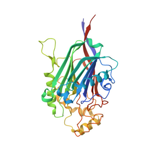
5A7J | pdb_00005a7j

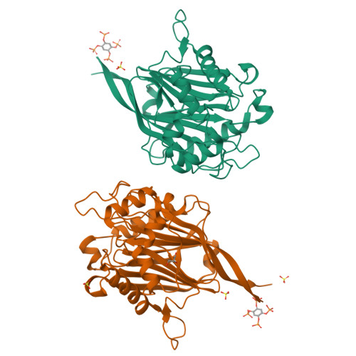
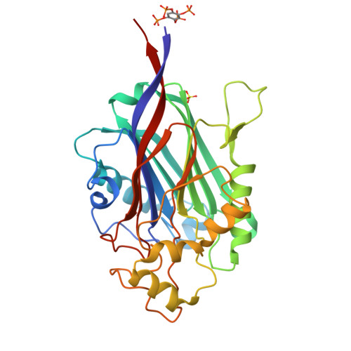
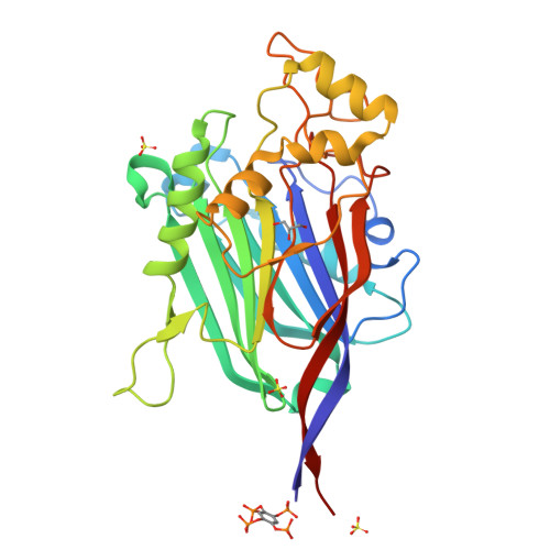
Crystal structure of INPP5B in complex with benzene 1,2,4,5- tetrakisphosphate


Experimental Data Snapshot

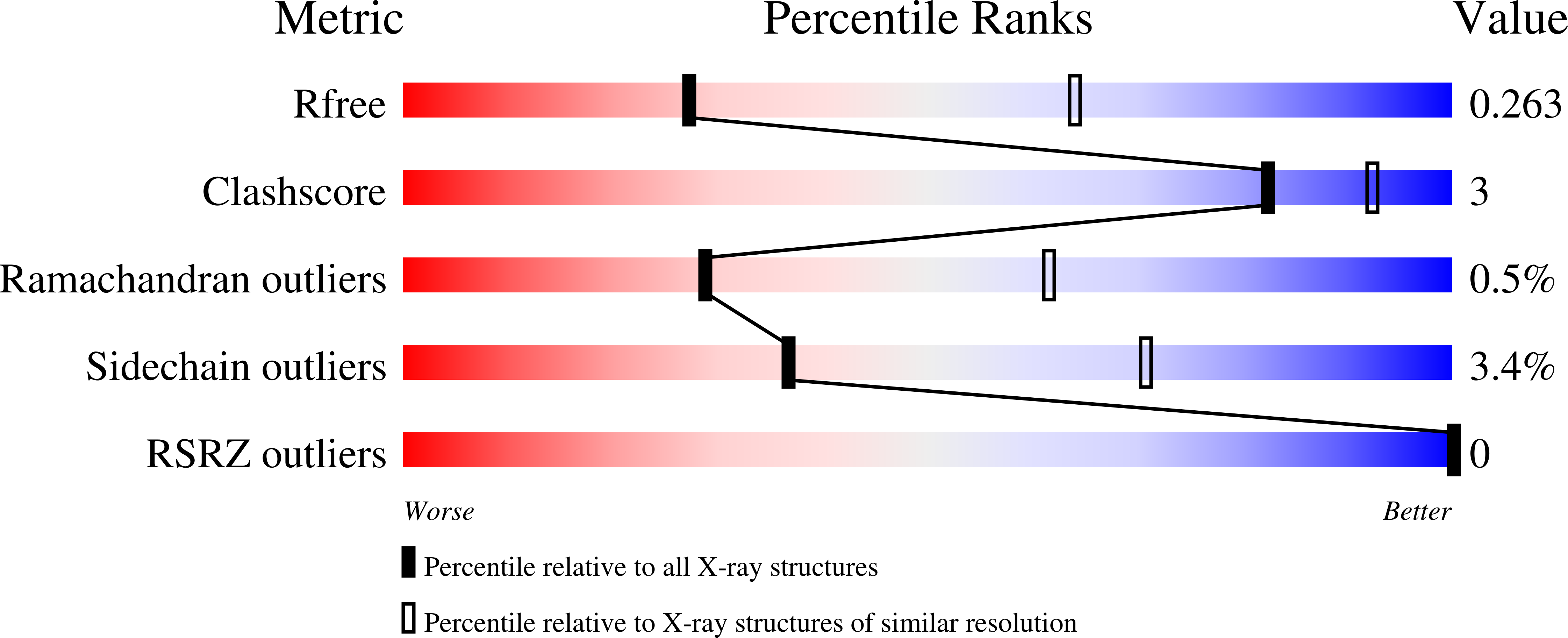
  • Method: X-RAY DIFFRACTION
  • Resolution: 2.90 Å
  • R-Value Free: 
    0.250 (Depositor), 0.260 (DCC) 
  • R-Value Work: 
    0.200 (Depositor), 0.210 (DCC) 
  • R-Value Observed: 
    0.202 (Depositor) 

Starting Model: experimental
View more details

wwPDB Validation   3D Report Full Report


Ligand Structure Quality Assessment 

Created with Raphaël 2.3.0Worse 01 BetterLigand structure goodness of fit to experimental dataBest fitted K2YClick on this verticalbar to view details

This is version 2.1 of the entry. See complete history


Literature

Crystal Structures of Type-II Inositol Polyphosphate 5-Phosphatase Inpp5B with Synthetic Inositol Polyphosphate Surrogates Reveal New Mechanistic Insights for the Inositol 5-Phosphatase Family.

Mills, S.J.Silvander, C.Cozier, G.Tresaugues, L.Nordlund, P.Potter, B.V.L.

(2016) Biochemistry 55: 1384

  • DOI: https://doi.org/10.1021/acs.biochem.5b00838
  • Primary Citation of Related Structures:  
    5A7I, 5A7J

  • PubMed Abstract: 

    The inositol polyphosphate 5-phosphatase INPP5B hydrolyzes the 5-phosphate group from water- and lipid-soluble signaling messengers. Two synthetic benzene and biphenyl polyphosphates (BzP/BiPhPs), simplified surrogates of inositol phosphates and phospholipid headgroups, were identified by thermodynamic studies as potent INPP5B ligands. The X-ray structure of the complex between INPP5B and biphenyl 3,3',4,4',5,5'-hexakisphosphate [BiPh(3,3',4,4',5,5')P6, IC50 5.5 μM] was determined at 2.89 Å resolution. One inhibitor pole locates in the phospholipid headgroup binding site and the second solvent-exposed ring binds to the His-Tag of another INPP5B molecule, while a molecule of inorganic phosphate is also present in the active site. Benzene 1,2,3-trisphosphate [Bz(1,2,3)P3] [one ring of BiPh(3,3',4,4',5,5')P6] inhibits INPP5B ca. 6-fold less potently. Co-crystallization with benzene 1,2,4,5-tetrakisphosphate [Bz(1,2,4,5)P4, IC50 = 6.3 μM] yielded a structure refined at 2.9 Å resolution. Conserved residues among the 5-phosphatase family mediate interactions with Bz(1,2,4,5)P4 and BiPh(3,3',4,4',5,5')P6 similar to those with the polar groups present in positions 1, 4, 5, and 6 on the inositol ring of the substrate. 5-Phosphatase specificity most likely resides in the variable zone located close to the 2- and 3-positions of the inositol ring, offering insights to inhibitor design. We propose that the inorganic phosphate present in the INPP5B-BiPh(3,3',4,4',5,5')P6 complex mimics the postcleavage substrate 5-phosphate released by INPP5B in the catalytic site, allowing elucidation of two new key features in the catalytic mechanism proposed for the family of phosphoinositide 5-phosphatases: first, the involvement of the conserved Arg-451 in the interaction with the 5-phosphate and second, identification of the water molecule that initiates 5-phosphate hydrolysis. Our model also has implications for the proposed "moving metal" mechanism.


  • Organizational Affiliation

    Wolfson Laboratory of Medicinal Chemistry, Department of Pharmacy and Pharmacology, University of Bath , Bath BA2 7AY, U.K.


Macromolecules
Find similar proteins by:  (by identity cutoff)  |  3D Structure
Entity ID: 1
MoleculeChains Sequence LengthOrganismDetailsImage
TYPE II INOSITOL 1,4,5-TRISPHOSPHATE 5-PHOSPHATASE
A, B
313Homo sapiensMutation(s): 0 
EC: 3.1.3.36
UniProt & NIH Common Fund Data Resources
Find proteins for P32019 (Homo sapiens)
Explore P32019 
Go to UniProtKB:  P32019
PHAROS:  P32019
GTEx:  ENSG00000204084 
Entity Groups  
Sequence Clusters30% Identity50% Identity70% Identity90% Identity95% Identity100% Identity
UniProt GroupP32019
Sequence Annotations
Expand
  • Reference Sequence
Small Molecules
Ligands 3 Unique
IDChains Name / Formula / InChI Key2D Diagram3D Interactions
K2Y
Query on K2Y

Download Ideal Coordinates CCD File 
D [auth A],
H [auth B]
(2,4,5-triphosphonooxyphenyl) dihydrogen phosphate
C6 H10 O16 P4
OFSQKFMVBPORNX-UHFFFAOYSA-N
SO4
Query on SO4

Download Ideal Coordinates CCD File 
C [auth A],
F [auth B],
G [auth B],
I [auth B]
SULFATE ION
O4 S
QAOWNCQODCNURD-UHFFFAOYSA-L
GOL
Query on GOL

Download Ideal Coordinates CCD File 
E [auth B]GLYCEROL
C3 H8 O3
PEDCQBHIVMGVHV-UHFFFAOYSA-N
Binding Affinity Annotations 
IDSourceBinding Affinity
K2Y BindingDB:  5A7J IC50: 6300 (nM) from 1 assay(s)
Experimental Data & Validation

Experimental Data

  • Method: X-RAY DIFFRACTION
  • Resolution: 2.90 Å
  • R-Value Free:  0.250 (Depositor), 0.260 (DCC) 
  • R-Value Work:  0.200 (Depositor), 0.210 (DCC) 
  • R-Value Observed: 0.202 (Depositor) 
Space Group: P 1 21 1
Unit Cell:
Length ( Å )Angle ( ˚ )
a = 52.261α = 90
b = 94.442β = 106.32
c = 78.313γ = 90
Software Package:
Software NamePurpose
BUSTER-TNTrefinement
XDSdata reduction
XDSdata scaling
PHASERphasing

Structure Validation

View Full Validation Report


Ligand Structure Quality Assessment 

Created with Raphaël 2.3.0Worse 01 BetterLigand structure goodness of fit to experimental dataBest fitted K2YClick on this verticalbar to view details

Entry History 

Deposition Data

Revision History  (Full details and data files)

  • Version 1.0: 2016-04-13
    Type: Initial release
  • Version 2.0: 2018-10-03
    Changes: Atomic model, Data collection, Derived calculations, Structure summary
  • Version 2.1: 2024-01-10
    Changes: Data collection, Database references, Derived calculations, Other, Refinement description