5YCG

Ancestral myoglobin aMbWp of Pakicetus relative


Experimental Data Snapshot

  • Method: X-RAY DIFFRACTION
  • Resolution: 2.40 Å
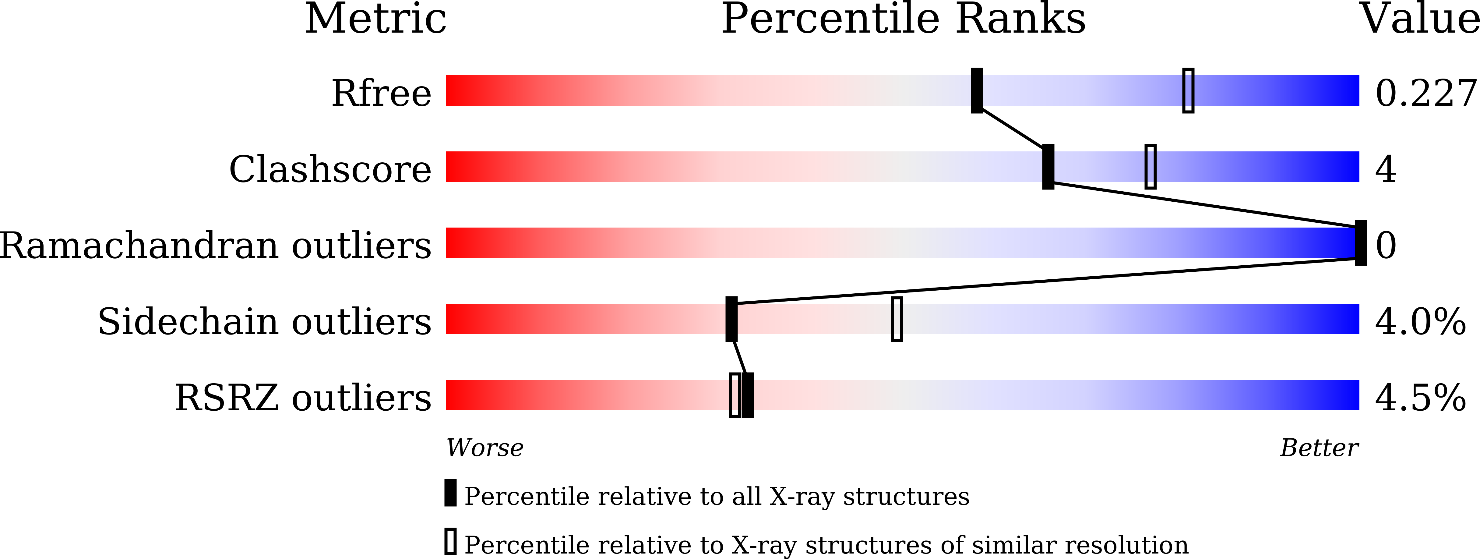
  • R-Value Free: 0.227 
  • R-Value Work: 0.184 
  • R-Value Observed: 0.186 

Starting Model: experimental
View more details

wwPDB Validation   3D Report Full Report


Ligand Structure Quality Assessment 


This is version 1.2 of the entry. See complete history


Literature

Tracing whale myoglobin evolution by resurrecting ancient proteins.

Isogai, Y.Imamura, H.Nakae, S.Sumi, T.Takahashi, K.I.Nakagawa, T.Tsuneshige, A.Shirai, T.

(2018) Sci Rep 8: 16883-16883

  • DOI: https://doi.org/10.1038/s41598-018-34984-6
  • Primary Citation of Related Structures:  
    5YCE, 5YCG, 5YCH, 5YCI, 5YCJ

  • PubMed Abstract: 

    Extant cetaceans, such as sperm whale, acquired the great ability to dive into the ocean depths during the evolution from their terrestrial ancestor that lived about 50 million years ago. Myoglobin (Mb) is highly concentrated in the myocytes of diving animals, in comparison with those of land animals, and is thought to play a crucial role in their adaptation as the molecular aqualung. Here, we resurrected ancestral whale Mbs, which are from the common ancestor between toothed and baleen whales (Basilosaurus), and from a further common quadrupedal ancestor between whale and hippopotamus (Pakicetus). The experimental and theoretical analyses demonstrated that whale Mb adopted two distinguished strategies to increase the protein concentration in vivo along the evolutionary history of deep sea adaptation; gaining precipitant tolerance in the early phase of the evolution, and increase of folding stability in the late phase.


  • Organizational Affiliation

    Department of Pharmaceutical Engineering, Toyama Prefectural University, Imizu, Toyama, 939-0398, Japan. [email protected].


Macromolecules
Find similar proteins by:  (by identity cutoff)  |  3D Structure
Entity ID: 1
MoleculeChains Sequence LengthOrganismDetailsImage
Ancestral myoglobin aMbWp of Pakicetus relative157Physeter macrocephalusMutation(s): 0 
EC: 1.11.1 (UniProt), 1.7 (UniProt)
UniProt
Find proteins for P68082 (Equus caballus)
Explore P68082 
Go to UniProtKB:  P68082
Entity Groups  
Sequence Clusters30% Identity50% Identity70% Identity90% Identity95% Identity100% Identity
UniProt GroupP68082
Sequence Annotations
Expand
  • Reference Sequence
Small Molecules
Ligands 1 Unique
IDChains Name / Formula / InChI Key2D Diagram3D Interactions
HEM
Query on HEM

Download Ideal Coordinates CCD File 
B [auth A]PROTOPORPHYRIN IX CONTAINING FE
C34 H32 Fe N4 O4
KABFMIBPWCXCRK-RGGAHWMASA-L
Experimental Data & Validation

Experimental Data

  • Method: X-RAY DIFFRACTION
  • Resolution: 2.40 Å
  • R-Value Free: 0.227 
  • R-Value Work: 0.184 
  • R-Value Observed: 0.186 
  • Space Group: P 1 21 1
Unit Cell:
Length ( Å )Angle ( ˚ )
a = 55.39α = 90
b = 29.74β = 112.65
c = 58.36γ = 90
Software Package:
Software NamePurpose
PHENIXrefinement
iMOSFLMdata reduction
SCALAdata scaling
PHENIXphasing

Structure Validation

View Full Validation Report


Ligand Structure Quality Assessment 


Entry History & Funding Information

Deposition Data


Funding OrganizationLocationGrant Number
Ministry of Education, Culture, Sports, Science and Technology (Japan)JapanJP17H01818
Japan Agency for Medical Research and DevelopmentJapan17am0101069j0001

Revision History  (Full details and data files)

  • Version 1.0: 2018-09-19
    Type: Initial release
  • Version 1.1: 2018-12-12
    Changes: Data collection, Database references
  • Version 1.2: 2023-11-22
    Changes: Data collection, Database references, Refinement description