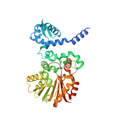
4WXH

Carminomycin-4-O-methyltransferase (DnrK) variant (298Ser insert) in complex with S-adenosyl-L-homocysteine (SAH) and aclacinomycin T


Experimental Data Snapshot

  • Method: X-RAY DIFFRACTION
  • Resolution: 1.90 Å
  • R-Value Free: 0.234 
  • R-Value Work: 0.193 
  • R-Value Observed: 0.195 

Starting Model: experimental
View more details

wwPDB Validation   3D Report Full Report


Ligand Structure Quality Assessment 


This is version 1.4 of the entry. See complete history


Literature

Divergent evolution of an atypical S-adenosyl-l-methionine-dependent monooxygenase involved in anthracycline biosynthesis.

Grocholski, T.Dinis, P.Niiranen, L.Niemi, J.Metsa-Ketela, M.

(2015) Proc Natl Acad Sci U S A 112: 9866-9871

  • DOI: https://doi.org/10.1073/pnas.1501765112
  • Primary Citation of Related Structures:  
    4WXH

  • PubMed Abstract: 

    Bacterial secondary metabolic pathways are responsible for the biosynthesis of thousands of bioactive natural products. Many enzymes residing in these pathways have evolved to catalyze unusual chemical transformations, which is facilitated by an evolutionary pressure promoting chemical diversity. Such divergent enzyme evolution has been observed in S-adenosyl-L-methionine (SAM)-dependent methyltransferases involved in the biosynthesis of anthracycline anticancer antibiotics; whereas DnrK from the daunorubicin pathway is a canonical 4-O-methyltransferase, the closely related RdmB (52% sequence identity) from the rhodomycin pathways is an atypical 10-hydroxylase that requires SAM, a thiol reducing agent, and molecular oxygen for activity. Here, we have used extensive chimeragenesis to gain insight into the functional differentiation of RdmB and show that insertion of a single serine residue to DnrK is sufficient for introduction of the monooxygenation activity. The crystal structure of DnrK-Ser in complex with aclacinomycin T and S-adenosyl-L-homocysteine refined to 1.9-Å resolution revealed that the inserted serine S297 resides in an α-helical segment adjacent to the substrate, but in a manner where the side chain points away from the active site. Further experimental work indicated that the shift in activity is mediated by rotation of a preceding phenylalanine F296 toward the active site, which blocks a channel to the surface of the protein that is present in native DnrK. The channel is also closed in RdmB and may be important for monooxygenation in a solvent-free environment. Finally, we postulate that the hydroxylation ability of RdmB originates from a previously undetected 10-decarboxylation activity of DnrK.


  • Organizational Affiliation

    Department of Biochemistry, University of Turku, FIN-20014 Turku, Finland.


Macromolecules
Find similar proteins by:  (by identity cutoff)  |  3D Structure
Entity ID: 1
MoleculeChains Sequence LengthOrganismDetailsImage
Carminomycin 4-O-methyltransferase DnrK
A, B
367Streptomyces peucetiusMutation(s): 0 
Gene Names: dnrK
EC: 2.1.1.292
UniProt
Find proteins for Q06528 (Streptomyces peucetius)
Explore Q06528 
Go to UniProtKB:  Q06528
Entity Groups  
Sequence Clusters30% Identity50% Identity70% Identity90% Identity95% Identity100% Identity
UniProt GroupQ06528
Sequence Annotations
Expand
  • Reference Sequence
Small Molecules
Ligands 4 Unique
IDChains Name / Formula / InChI Key2D Diagram3D Interactions
3VL
Query on 3VL

Download Ideal Coordinates CCD File 
D [auth A],
J [auth B]
methyl (1R,2R,4S)-2-ethyl-2,5,7-trihydroxy-6,11-dioxo-4-{[2,3,6-trideoxy-3-(dimethylamino)-alpha-L-lyxo-hexopyranosyl]oxy}-1,2,3,4,6,11-hexahydrotetracene-1-carboxylate
C30 H35 N O10
LJZPVWKMAYDYAS-QKKPTTNWSA-N
SAH
Query on SAH

Download Ideal Coordinates CCD File 
C [auth A],
I [auth B]
S-ADENOSYL-L-HOMOCYSTEINE
C14 H20 N6 O5 S
ZJUKTBDSGOFHSH-WFMPWKQPSA-N
DTU
Query on DTU

Download Ideal Coordinates CCD File 
E [auth A](2R,3S)-1,4-DIMERCAPTOBUTANE-2,3-DIOL
C4 H10 O2 S2
VHJLVAABSRFDPM-ZXZARUISSA-N
SO4
Query on SO4

Download Ideal Coordinates CCD File 
F [auth A],
G [auth A],
H [auth A],
K [auth B],
L [auth B]
SULFATE ION
O4 S
QAOWNCQODCNURD-UHFFFAOYSA-L
Experimental Data & Validation

Experimental Data

  • Method: X-RAY DIFFRACTION
  • Resolution: 1.90 Å
  • R-Value Free: 0.234 
  • R-Value Work: 0.193 
  • R-Value Observed: 0.195 
  • Space Group: P 1 21 1
Unit Cell:
Length ( Å )Angle ( ˚ )
a = 60.803α = 90
b = 100.995β = 102.47
c = 62.398γ = 90
Software Package:
Software NamePurpose
PHENIXrefinement
XSCALEdata scaling
MOLREPphasing
XDSdata reduction

Structure Validation

View Full Validation Report


Ligand Structure Quality Assessment 


Entry History & Funding Information

Deposition Data


Funding OrganizationLocationGrant Number
Academy of FinlandFinland136060

Revision History  (Full details and data files)

  • Version 1.0: 2015-07-29
    Type: Initial release
  • Version 1.1: 2015-08-12
    Changes: Database references
  • Version 1.2: 2015-08-19
    Changes: Database references
  • Version 1.3: 2018-02-28
    Changes: Structure summary
  • Version 1.4: 2024-01-10
    Changes: Data collection, Database references, Refinement description