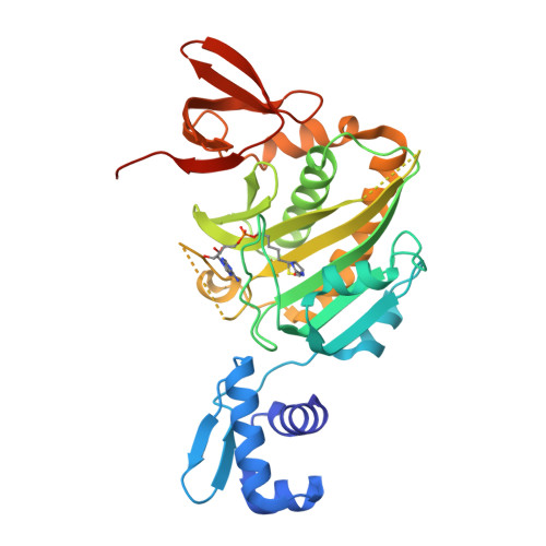
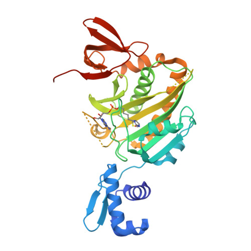
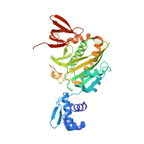
4WF2 | pdb_00004wf2

Structure of E. coli BirA G142A bound to biotinol-5'-AMP


Experimental Data Snapshot

  • Method: X-RAY DIFFRACTION
  • Resolution: 2.31 Å
  • R-Value Free: 
    0.218 (Depositor), 0.220 (DCC) 
  • R-Value Work: 
    0.172 (Depositor), 0.180 (DCC) 
  • R-Value Observed: 
    0.174 (Depositor) 

wwPDB Validation   3D Report Full Report


Ligand Structure Quality Assessment 

Created with Raphaël 2.3.0Worse 01 BetterLigand structure goodness of fit to experimental dataBest fitted BTXClick on this verticalbar to view details

This is version 1.6 of the entry. See complete history


Literature

Allosteric Coupling via Distant Disorder-to-Order Transitions.

Eginton, C.Cressman, W.J.Bachas, S.Wade, H.Beckett, D.

(2015) J Mol Biology 427: 1695-1704

  • DOI: https://doi.org/10.1016/j.jmb.2015.02.021
  • Primary Citation of Related Structures:  
    4WF2

  • PubMed Abstract: 

    Intrinsic disorder provides a means of maximizing allosteric coupling in proteins. However, the mechanisms by which the disorder functions in allostery remain to be elucidated. Small ligand, bio-5'-AMP, binding and dimerization of the Escherichia coli biotin repressor are allosterically coupled. Folding of a disordered loop in the allosteric effector binding site is required to realize the full coupling free energy of -4.0 ± 0.3 kcal/mol observed in the wild-type protein. Alanine substitution of a glycine residue on the dimerization surface that does not directly contribute to the dimerization interface completely abolishes this coupling. In this work, the structure of this variant, solved by X-ray crystallography, reveals a monomeric corepressor-bound protein. In the structure loops, neither of which contains the alanine substitution, on both the dimerization and effector binding surfaces that are folded in the corepressor-bound wild-type protein are disordered. The structural data combined with functional measurements indicate that allosteric coupling between ligand binding and dimerization in BirA (E. coli biotin repressor/biotin protein ligase) is achieved via reciprocal communication of disorder-to-order transitions on two distant functional surfaces.


  • Organizational Affiliation

    Department of Chemistry and Biochemistry, University of Maryland, College Park, MD 20742, USA.


Macromolecules
Find similar proteins by:  (by identity cutoff)  |  3D Structure
Entity ID: 1
MoleculeChains Sequence LengthOrganismDetailsImage
Bifunctional ligase/repressor BirA327Escherichia coli K-12Mutation(s): 1 
Gene Names: birAbioRdhbBb3973JW3941
EC: 6.3.4.15
UniProt
Find proteins for P06709 (Escherichia coli (strain K12))
Explore P06709 
Go to UniProtKB:  P06709
Entity Groups  
Sequence Clusters30% Identity50% Identity70% Identity90% Identity95% Identity100% Identity
UniProt GroupP06709
Sequence Annotations
Expand
  • Reference Sequence
Small Molecules
Ligands 1 Unique
IDChains Name / Formula / InChI Key2D Diagram3D Interactions
BTX
Query on BTX

Download Ideal Coordinates CCD File 
B [auth A]((2R,3S,4R,5R)-5-(6-AMINO-9H-PURIN-9-YL)-3,4-DIHYDROXY-TETRAHYDROFURAN-2-YL)METHYL 5-((3AS,4S,6AR)-2-OXO-HEXAHYDRO-1H-THIENO[3,4-D]IMIDAZOL-4-YL)PENTYL HYDROGEN PHOSPHATE
C20 H30 N7 O8 P S
KBOGUFFJCBPJEH-SQGSUPJISA-N
Binding Affinity Annotations 
IDSourceBinding Affinity
BTX BindingDB:  4WF2 Ki: 87 (nM) from 1 assay(s)
Kd: 1.5 (nM) from 1 assay(s)
Experimental Data & Validation

Experimental Data

  • Method: X-RAY DIFFRACTION
  • Resolution: 2.31 Å
  • R-Value Free:  0.218 (Depositor), 0.220 (DCC) 
  • R-Value Work:  0.172 (Depositor), 0.180 (DCC) 
  • R-Value Observed: 0.174 (Depositor) 
Space Group: P 43
Unit Cell:
Length ( Å )Angle ( ˚ )
a = 46.239α = 90
b = 46.239β = 90
c = 157.107γ = 90
Software Package:
Software NamePurpose
PHENIXrefinement

Structure Validation

View Full Validation Report


Ligand Structure Quality Assessment 

Created with Raphaël 2.3.0Worse 01 BetterLigand structure goodness of fit to experimental dataBest fitted BTXClick on this verticalbar to view details

Entry History & Funding Information

Deposition Data


Funding OrganizationLocationGrant Number
National Science Foundation (NSF, United States)United StatesMCB-0953430

Revision History  (Full details and data files)

  • Version 1.0: 2014-10-22
    Type: Initial release
  • Version 1.1: 2015-02-04
    Changes: Derived calculations
  • Version 1.2: 2015-03-25
    Changes: Database references
  • Version 1.3: 2015-04-08
    Changes: Database references
  • Version 1.4: 2017-09-27
    Changes: Author supporting evidence, Database references, Derived calculations, Other, Source and taxonomy, Structure summary
  • Version 1.5: 2019-11-27
    Changes: Author supporting evidence
  • Version 1.6: 2023-12-27
    Changes: Data collection, Database references, Refinement description