Structural and functional studies of conserved nucleotide-binding protein LptB in lipopolysaccharide transport.
Wang, Z., Xiang, Q., Zhu, X., Dong, H., He, C., Wang, H., Zhang, Y., Wang, W., Dong, C.(2014) Biochem Biophys Res Commun 452: 443-449
- PubMed: 25172661
- DOI: https://doi.org/10.1016/j.bbrc.2014.08.094
- Primary Citation of Related Structures:
4QC2 - PubMed Abstract:
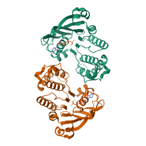
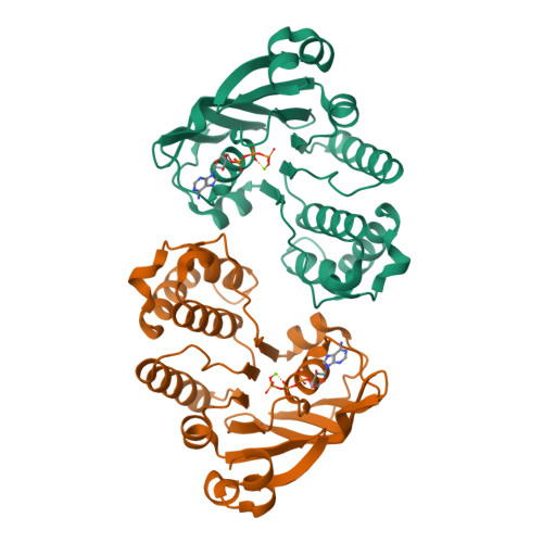
Lipopolysaccharide (LPS) is the main component of the outer membrane of Gram-negative bacteria, which plays an essential role in protecting the bacteria from harsh conditions and antibiotics. LPS molecules are transported from the inner membrane to the outer membrane by seven LPS transport proteins. LptB is vital in hydrolyzing ATP to provide energy for LPS transport, however this mechanism is not very clear. Here we report wild-type LptB crystal structure in complex with ATP and Mg(2+), which reveals that its structure is conserved with other nucleotide-binding proteins (NBD). Structural, functional and electron microscopic studies demonstrated that the ATP binding residues, including K42 and T43, are crucial for LptB's ATPase activity, LPS transport and the vitality of Escherichia coli cells with the exceptions of H195A and Q85A; the H195A mutation does not lower its ATPase activity but impairs LPS transport, and Q85A does not alter ATPase activity but causes cell death. Our data also suggest that two protomers of LptB have to work together for ATP hydrolysis and LPS transport. These results have significant impacts in understanding the LPS transport mechanism and developing new antibiotics.
Organizational Affiliation:
Biomedical Research Centre, Norwich Medical School, University of East Anglia, Norwich Research Park, NR4 7TJ, UK; College of Life Sciences, Sichuan University, Chengdu 610065, China; Biomedical Sciences Research Complex, School of Chemistry, University of St Andrews, North Haugh, St Andrews KY16 9ST, UK.


