X-ray-induced changes in the active site structure of octaheme cytochrome c nitrite reductase and its substrate complexes
Lazarenko, V.A., Polyakov, K.M., Trofimov, A.A., Popov, A.N., Tikhonova, T.V., Tikhonov, A.V., Popov, V.O.To be published.
Experimental Data Snapshot
Entity ID: 1 | |||||
|---|---|---|---|---|---|
| Molecule | Chains | Sequence Length | Organism | Details | Image |
| Eight-heme nitrite reductase | 521 | Thioalkalivibrio nitratireducens | Mutation(s): 0 EC: 1.7.2.2 | 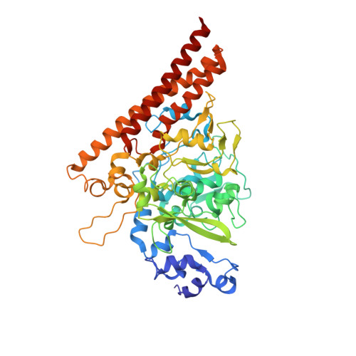 | |
UniProt | |||||
Find proteins for L0DSL2 (Thioalkalivibrio nitratireducens (strain DSM 14787 / UNIQEM 213 / ALEN2)) Explore L0DSL2 Go to UniProtKB: L0DSL2 | |||||
Entity Groups | |||||
| Sequence Clusters | 30% Identity50% Identity70% Identity90% Identity95% Identity100% Identity | ||||
| UniProt Group | L0DSL2 | ||||
Sequence AnnotationsExpand | |||||
| |||||
| Ligands 6 Unique | |||||
|---|---|---|---|---|---|
| ID | Chains | Name / Formula / InChI Key | 2D Diagram | 3D Interactions | |
| HEC Query on HEC | C [auth A] D [auth A] E [auth A] F [auth A] G [auth A] | HEME C C34 H34 Fe N4 O4 HXQIYSLZKNYNMH-LJNAALQVSA-N |  | ||
| TRS Query on TRS | GA [auth B], P [auth A] | 2-AMINO-2-HYDROXYMETHYL-PROPANE-1,3-DIOL C4 H12 N O3 LENZDBCJOHFCAS-UHFFFAOYSA-O |  | ||
| MPD Query on MPD | DA [auth B], M [auth A] | (4S)-2-METHYL-2,4-PENTANEDIOL C6 H14 O2 SVTBMSDMJJWYQN-YFKPBYRVSA-N |  | ||
| SO3 Query on SO3 | BA [auth B], EA [auth B], K [auth A], N [auth A], Q [auth A] | SULFITE ION O3 S LSNNMFCWUKXFEE-UHFFFAOYSA-L |  | ||
| ACT Query on ACT | AA [auth B], FA [auth B], O [auth A], R [auth A] | ACETATE ION C2 H3 O2 QTBSBXVTEAMEQO-UHFFFAOYSA-M |  | ||
| CA Query on CA | CA [auth B], L [auth A] | CALCIUM ION Ca BHPQYMZQTOCNFJ-UHFFFAOYSA-N |  | ||
| Length ( Å ) | Angle ( ˚ ) |
|---|---|
| a = 195.2 | α = 90 |
| b = 195.2 | β = 90 |
| c = 195.2 | γ = 90 |
| Software Name | Purpose |
|---|---|
| REFMAC | refinement |
| XDS | data reduction |
| XSCALE | data scaling |
RCSB PDB is hosted by

RCSB PDB is a member of the