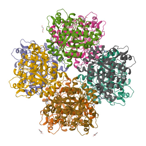
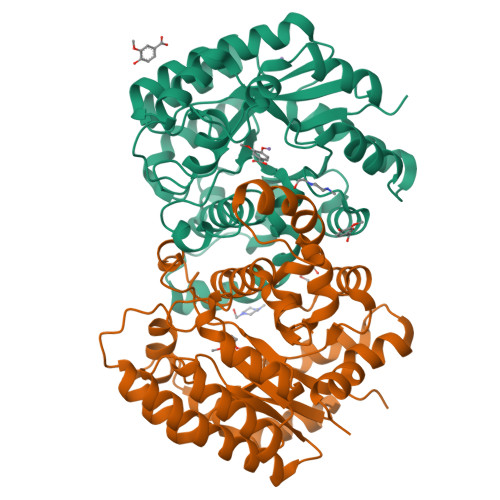
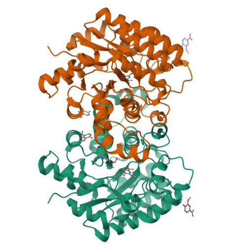
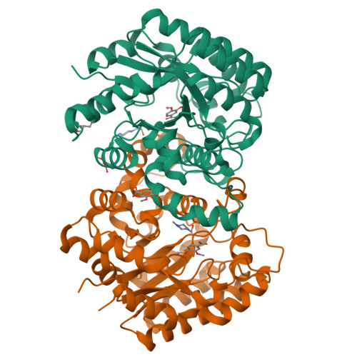
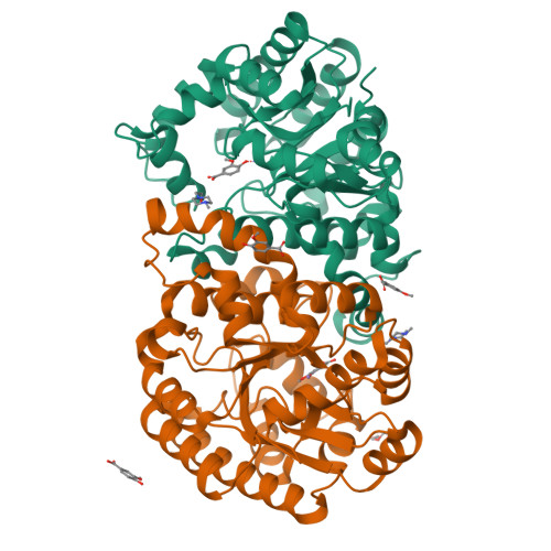
Crystal structure of 5-carboxyvanillate decarboxylase from Sphingomonas paucimobilis complexed with vanillic acid
Fedorov, A.A., Fedorov, E.V., Vladimirova, A., Raushel, F.M., Almo, S.C.To be published.
Experimental Data Snapshot
Starting Model: experimental
View more details
Entity ID: 1 | |||||
|---|---|---|---|---|---|
| Molecule | Chains | Sequence Length | Organism | Details | Image |
| 5-carboxyvanillate decarboxylase | 335 | Sphingomonas paucimobilis | Mutation(s): 0 Gene Names: ligW | 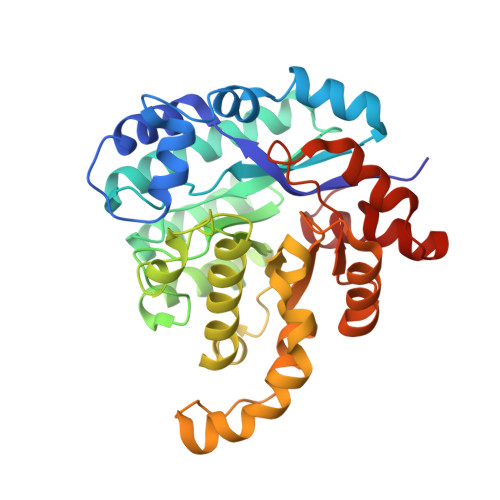 | |
UniProt | |||||
Find proteins for Q8RJ47 (Sphingomonas paucimobilis) Explore Q8RJ47 Go to UniProtKB: Q8RJ47 | |||||
Entity Groups | |||||
| Sequence Clusters | 30% Identity50% Identity70% Identity90% Identity95% Identity100% Identity | ||||
| UniProt Group | Q8RJ47 | ||||
Sequence AnnotationsExpand | |||||
| |||||
| Ligands 5 Unique | |||||
|---|---|---|---|---|---|
| ID | Chains | Name / Formula / InChI Key | 2D Diagram | 3D Interactions | |
| EPE Query on EPE | BA [auth D] GA [auth E] LA [auth F] M [auth A] QA [auth G] | 4-(2-HYDROXYETHYL)-1-PIPERAZINE ETHANESULFONIC ACID C8 H18 N2 O4 S JKMHFZQWWAIEOD-UHFFFAOYSA-N |  | ||
| VNL Query on VNL | AA [auth D] DA [auth E] EA [auth E] FA [auth E] J [auth A] | 4-HYDROXY-3-METHOXYBENZOATE C8 H7 O4 WKOLLVMJNQIZCI-UHFFFAOYSA-M |  | ||
| PEG Query on PEG | HA [auth E] | DI(HYDROXYETHYL)ETHER C4 H10 O3 MTHSVFCYNBDYFN-UHFFFAOYSA-N |  | ||
| GOL Query on GOL | X [auth C] | GLYCEROL C3 H8 O3 PEDCQBHIVMGVHV-UHFFFAOYSA-N |  | ||
| MN Query on MN | CA [auth E] I [auth A] IA [auth F] MA [auth G] N [auth A] | MANGANESE (II) ION Mn WAEMQWOKJMHJLA-UHFFFAOYSA-N |  | ||
| Length ( Å ) | Angle ( ˚ ) |
|---|---|
| a = 80.592 | α = 109.71 |
| b = 96.917 | β = 90.53 |
| c = 97.029 | γ = 111.89 |
| Software Name | Purpose |
|---|---|
| CBASS | data collection |
| BALBES | phasing |
| PHENIX | refinement |
| HKL-2000 | data reduction |
| HKL-2000 | data scaling |