4HUN | pdb_00004hun

MATE transporter NorM-NG in complex with R6G and monobody


Experimental Data Snapshot

  • Method: X-RAY DIFFRACTION
  • Resolution: 3.59 Å
  • R-Value Free: 
    0.327 (Depositor), 0.340 (DCC) 
  • R-Value Work: 
    0.306 (Depositor), 0.320 (DCC) 
  • R-Value Observed: 
    0.307 (Depositor) 

wwPDB Validation   3D Report Full Report


Ligand Structure Quality Assessment 

Created with Raphaël 2.3.0Worse 01 BetterLigand structure goodness of fit to experimental dataBest fitted RHQClick on this verticalbar to view details

This is version 1.3 of the entry. See complete history


Literature

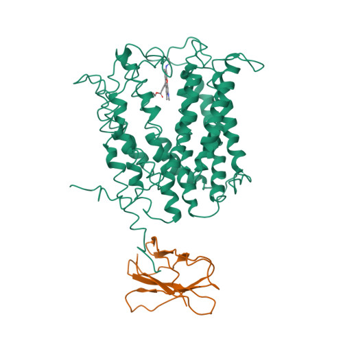
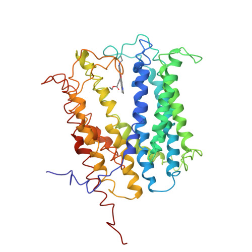
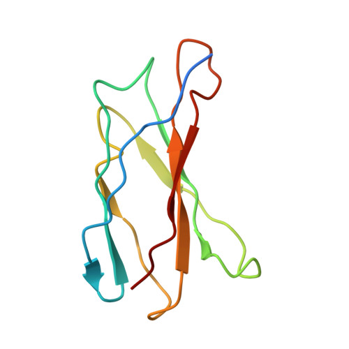
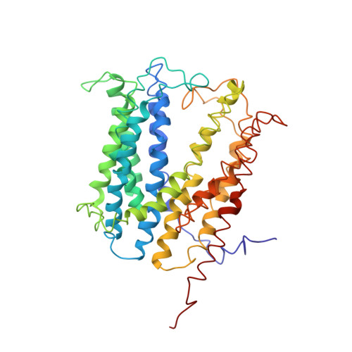
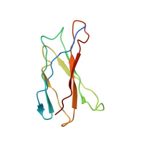
Structures of a Na+-coupled, substrate-bound MATE multidrug transporter.

Lu, M.Symersky, J.Radchenko, M.Koide, A.Guo, Y.Nie, R.Koide, S.

(2013) Proc Natl Acad Sci U S A 110: 2099-2104

  • DOI: https://doi.org/10.1073/pnas.1219901110
  • Primary Citation of Related Structures:  
    4HUK, 4HUL, 4HUM, 4HUN

  • PubMed Abstract: 

    Multidrug transporters belonging to the multidrug and toxic compound extrusion (MATE) family expel dissimilar lipophilic and cationic drugs across cell membranes by dissipating a preexisting Na(+) or H(+) gradient. Despite its clinical relevance, the transport mechanism of MATE proteins remains poorly understood, largely owing to a lack of structural information on the substrate-bound transporter. Here we report crystal structures of a Na(+)-coupled MATE transporter NorM from Neisseria gonorrheae in complexes with three distinct translocation substrates (ethidium, rhodamine 6G, and tetraphenylphosphonium), as well as Cs(+) (a Na(+) congener), all captured in extracellular-facing and drug-bound states. The structures revealed a multidrug-binding cavity festooned with four negatively charged amino acids and surprisingly limited hydrophobic moieties, in stark contrast to the general belief that aromatic amino acids play a prominent role in multidrug recognition. Furthermore, we discovered an uncommon cation-π interaction in the Na(+)-binding site located outside the drug-binding cavity and validated the biological relevance of both the substrate- and cation-binding sites by conducting drug resistance and transport assays. Additionally, we uncovered potential rearrangement of at least two transmembrane helices upon Na(+)-induced drug export. Based on our structural and functional analyses, we suggest that Na(+) triggers multidrug extrusion by inducing protein conformational changes rather than by directly competing for the substrate-binding amino acids. This scenario is distinct from the canonical antiport mechanism, in which both substrate and counterion compete for a shared binding site in the transporter. Collectively, our findings provide an important step toward a detailed and mechanistic understanding of multidrug transport.


  • Organizational Affiliation

    Department of Biochemistry and Molecular Biology, Rosalind Franklin University of Medicine and Science, North Chicago, IL 60064, USA. min.lu@rosalindfranklin.edu


Macromolecules
Find similar proteins by:  (by identity cutoff)  |  3D Structure
Entity ID: 1
MoleculeChains Sequence LengthOrganismDetailsImage
Multidrug efflux protein459Neisseria gonorrhoeae TCDC-NG08107Mutation(s): 0 
Gene Names: NGTW08_0430
Entity Groups  
Sequence Clusters30% Identity50% Identity70% Identity90% Identity95% Identity100% Identity
Sequence Annotations
Expand
  • Reference Sequence
Find similar proteins by:  (by identity cutoff)  |  3D Structure
Entity ID: 2
MoleculeChains Sequence LengthOrganismDetailsImage
protein B99Escherichia coliMutation(s): 0 
UniProt
Find proteins for M1E1G6 (Escherichia coli)
Explore M1E1G6 
Go to UniProtKB:  M1E1G6
Entity Groups  
Sequence Clusters30% Identity50% Identity70% Identity90% Identity95% Identity100% Identity
UniProt GroupM1E1G6
Sequence Annotations
Expand
  • Reference Sequence
Small Molecules
Ligands 1 Unique
IDChains Name / Formula / InChI Key2D Diagram3D Interactions
RHQ
Query on RHQ

Download Ideal Coordinates CCD File 
C [auth A]RHODAMINE 6G
C28 H31 N2 O3
IWWWBRIIGAXLCJ-KRUMMXJUSA-O
Experimental Data & Validation

Experimental Data

  • Method: X-RAY DIFFRACTION
  • Resolution: 3.59 Å
  • R-Value Free:  0.327 (Depositor), 0.340 (DCC) 
  • R-Value Work:  0.306 (Depositor), 0.320 (DCC) 
  • R-Value Observed: 0.307 (Depositor) 
Space Group: P 32 2 1
Unit Cell:
Length ( Å )Angle ( ˚ )
a = 117.234α = 90
b = 117.234β = 90
c = 226.218γ = 120
Software Package:
Software NamePurpose
HKL-2000data collection
SHARPphasing
REFMACrefinement
HKL-2000data reduction
HKL-2000data scaling

Structure Validation

View Full Validation Report


Ligand Structure Quality Assessment 

Created with Raphaël 2.3.0Worse 01 BetterLigand structure goodness of fit to experimental dataBest fitted RHQClick on this verticalbar to view details

Entry History 

Deposition Data

  • Released Date: 2013-02-06 
  • Deposition Author(s): Lu, M.

Revision History  (Full details and data files)

  • Version 1.0: 2013-02-06
    Type: Initial release
  • Version 1.1: 2013-03-06
    Changes: Database references
  • Version 1.2: 2021-04-28
    Changes: Advisory, Database references, Derived calculations
  • Version 1.3: 2024-02-28
    Changes: Data collection, Database references