4FR0

ArsM arsenic(III) S-adenosylmethionine methyltransferase with SAM


Experimental Data Snapshot

  • Method: X-RAY DIFFRACTION
  • Resolution: 2.75 Å
  • R-Value Free: 
    0.283 (Depositor), 0.280 (DCC) 
  • R-Value Work: 
    0.206 (Depositor), 0.200 (DCC) 
  • R-Value Observed: 
    0.209 (Depositor) 

Starting Model: experimental
View more details

wwPDB Validation   3D Report Full Report


Ligand Structure Quality Assessment 

Created with Raphaël 2.3.0Worse 01 BetterLigand structure goodness of fit to experimental dataBest fitted SAMClick on this verticalbar to view details

This is version 1.3 of the entry. See complete history


Literature

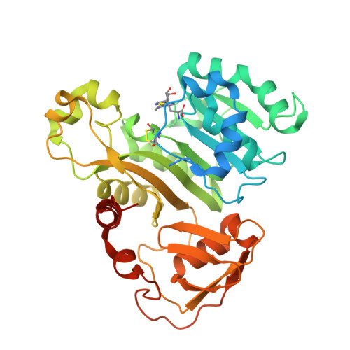
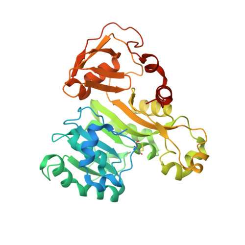
Structure of an As(III) S-Adenosylmethionine Methyltransferase: Insights into the Mechanism of Arsenic Biotransformation.

Ajees, A.A.Marapakala, K.Packianathan, C.Sankaran, B.Rosen, B.P.

(2012) Biochemistry 51: 5476-5485

  • DOI: https://doi.org/10.1021/bi3004632
  • Primary Citation of Related Structures:  
    4FR0, 4FS8, 4FSD

  • PubMed Abstract: 

    Enzymatic methylation of arsenic is a detoxification process in microorganisms but in humans may activate the metalloid to more carcinogenic species. We describe the first structure of an As(III) S-adenosylmethionine methyltransferase by X-ray crystallography that reveals a novel As(III) binding domain. The structure of the methyltransferase from the thermophilic eukaryotic alga Cyanidioschyzon merolae reveals the relationship between the arsenic and S-adenosylmethionine binding sites to a final resolution of ∼1.6 Å. As(III) binding causes little change in conformation, but binding of SAM reorients helix α4 and a loop (residues 49-80) toward the As(III) binding domain, positioning the methyl group for transfer to the metalloid. There is no evidence of a reductase domain. These results are consistent with previous suggestions that arsenic remains trivalent during the catalytic cycle. A homology model of human As(III) S-adenosylmethionine methyltransferase with the location of known polymorphisms was constructed. The structure provides insights into the mechanism of substrate binding and catalysis.


  • Organizational Affiliation

    Department of Cellular Biology and Pharmacology, Herbert Wertheim College of Medicine, Florida International University, Miami, 33199, United States.


Macromolecules
Find similar proteins by:  (by identity cutoff)  |  3D Structure
Entity ID: 1
MoleculeChains Sequence LengthOrganismDetailsImage
Arsenic methyltransferase383Cyanidioschyzon sp. 5508Mutation(s): 0 
Gene Names: arsM
EC: 2.1.1.137
UniProt
Find proteins for C0JV69 (Cyanidioschyzon sp. 5508)
Explore C0JV69 
Go to UniProtKB:  C0JV69
Entity Groups  
Sequence Clusters30% Identity50% Identity70% Identity90% Identity95% Identity100% Identity
UniProt GroupC0JV69
Sequence Annotations
Expand
  • Reference Sequence
Small Molecules
Ligands 1 Unique
IDChains Name / Formula / InChI Key2D Diagram3D Interactions
SAM
Query on SAM

Download Ideal Coordinates CCD File 
B [auth A]S-ADENOSYLMETHIONINE
C15 H22 N6 O5 S
MEFKEPWMEQBLKI-FCKMPRQPSA-N
Experimental Data & Validation

Experimental Data

  • Method: X-RAY DIFFRACTION
  • Resolution: 2.75 Å
  • R-Value Free:  0.283 (Depositor), 0.280 (DCC) 
  • R-Value Work:  0.206 (Depositor), 0.200 (DCC) 
  • R-Value Observed: 0.209 (Depositor) 
Space Group: C 2 2 2
Unit Cell:
Length ( Å )Angle ( ˚ )
a = 67.183α = 90
b = 128.789β = 90
c = 96.52γ = 90
Software Package:
Software NamePurpose
HKL-2000data collection
PHASERphasing
REFMACrefinement
HKL-2000data reduction
HKL-2000data scaling

Structure Validation

View Full Validation Report


Ligand Structure Quality Assessment 

Created with Raphaël 2.3.0Worse 01 BetterLigand structure goodness of fit to experimental dataBest fitted SAMClick on this verticalbar to view details

Entry History 

Deposition Data

Revision History  (Full details and data files)

  • Version 1.0: 2012-07-11
    Type: Initial release
  • Version 1.1: 2012-10-03
    Changes: Database references
  • Version 1.2: 2023-09-13
    Changes: Data collection, Database references, Derived calculations, Refinement description
  • Version 1.3: 2024-11-20
    Changes: Structure summary