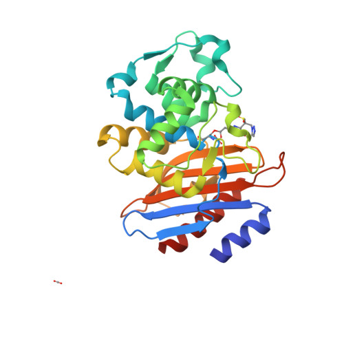
Crystal Structure of Class a Beta-Lactamase from Bacillus Licheniformis Inhibited by Tazobactam
Power, P., Herman, R., Kerff, F., Mercuri, P., Galleni, M., Gutkind, G., Charlier, P., Sauvage, E.To be published.
Experimental Data Snapshot
Starting Model: experimental
View more details
Entity ID: 1 | |||||
|---|---|---|---|---|---|
| Molecule | Chains | Sequence Length | Organism | Details | Image |
| BETA-LACTAMASE | 265 | Bacillus licheniformis | Mutation(s): 0 EC: 3.5.2.6 |  | |
UniProt | |||||
Find proteins for P94458 (Bacillus licheniformis) Explore P94458 Go to UniProtKB: P94458 | |||||
Entity Groups | |||||
| Sequence Clusters | 30% Identity50% Identity70% Identity90% Identity95% Identity100% Identity | ||||
| UniProt Group | P94458 | ||||
Sequence AnnotationsExpand | |||||
| |||||
| Ligands 4 Unique | |||||
|---|---|---|---|---|---|
| ID | Chains | Name / Formula / InChI Key | 2D Diagram | 3D Interactions | |
| TBE Query on TBE | C [auth A], G [auth B] | TAZOBACTAM INTERMEDIATE C10 H14 N4 O5 S ANZZKUOZZHRUQC-AARLMMRRSA-N |  | ||
| CIT Query on CIT | D [auth A] | CITRIC ACID C6 H8 O7 KRKNYBCHXYNGOX-UHFFFAOYSA-N |  | ||
| PGE Query on PGE | E [auth A] | TRIETHYLENE GLYCOL C6 H14 O4 ZIBGPFATKBEMQZ-UHFFFAOYSA-N |  | ||
| CO2 Query on CO2 | F [auth B], H [auth B] | CARBON DIOXIDE C O2 CURLTUGMZLYLDI-UHFFFAOYSA-N |  | ||
| Length ( Å ) | Angle ( ˚ ) |
|---|---|
| a = 46.955 | α = 90 |
| b = 103.55 | β = 94.77 |
| c = 63.732 | γ = 90 |
| Software Name | Purpose |
|---|---|
| REFMAC | refinement |
| MOSFLM | data reduction |
| SCALA | data scaling |