3U8X

Crystal Structure of a chimera containing the N-terminal domain (residues 8-29) of drosophila Ciboulot and the C-terminal domain (residues 18-44) of bovine Thymosin-beta4, bound to G-actin-ATP


Experimental Data Snapshot

  • Method: X-RAY DIFFRACTION
  • Resolution: 2.00 Å
  • R-Value Free: 
    0.238 (Depositor), 0.240 (DCC) 
  • R-Value Work: 
    0.186 (Depositor), 0.190 (DCC) 
  • R-Value Observed: 
    0.190 (Depositor) 

Starting Model: experimental
View more details

wwPDB Validation   3D Report Full Report


Ligand Structure Quality Assessment 

Created with Raphaël 2.3.0Worse 01 BetterLigand structure goodness of fit to experimental dataBest fitted ATPClick on this verticalbar to view details

This is version 1.3 of the entry. See complete history


Literature

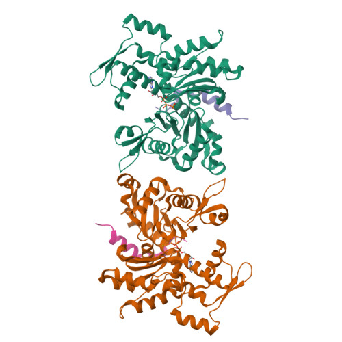
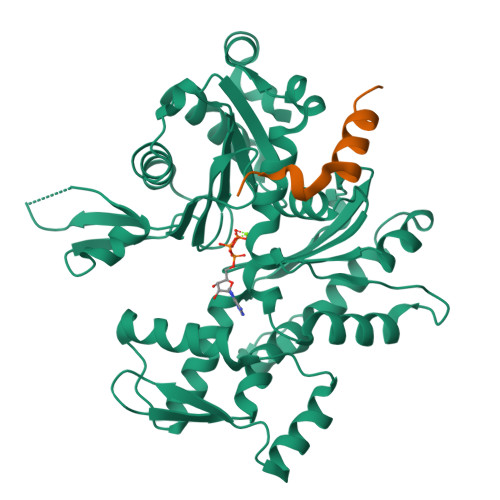
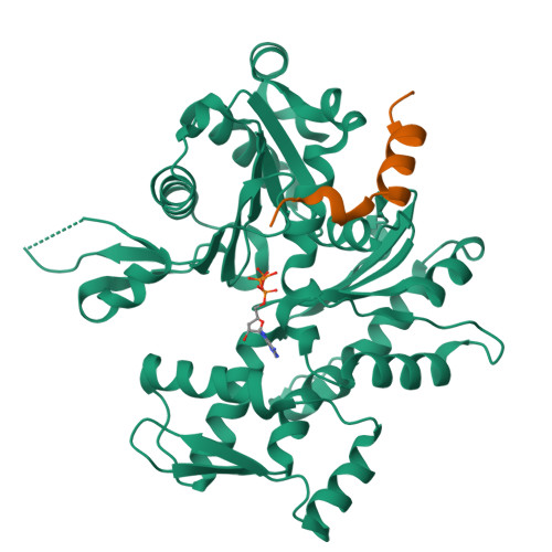
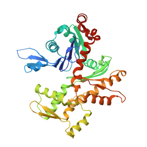
How a single residue in individual beta-thymosin/WH2 domains controls their functions in actin assembly

Didry, D.Cantrelle, F.X.Husson, C.Roblin, P.Moorthy, A.M.Perez, J.Le Clainche, C.Hertzog, M.Guittet, E.Carlier, M.F.van Heijenoort, C.Renault, L.

(2012) EMBO J 31: 1000-1013

  • DOI: https://doi.org/10.1038/emboj.2011.461
  • Primary Citation of Related Structures:  
    3SJH, 3U8X, 3U9D, 3U9Z

  • PubMed Abstract: 

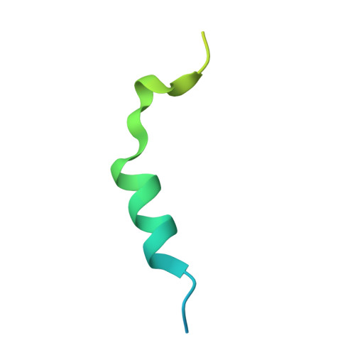
    β-Thymosin (βT) and WH2 domains are widespread, intrinsically disordered actin-binding peptides that display significant sequence variability and different regulations of actin self-assembly in motile and morphogenetic processes. Here, we reveal the structural mechanisms by which, in their 1:1 stoichiometric complexes with actin, they either inhibit assembly by sequestering actin monomers like Thymosin-β4, or enhance motility by directing polarized filament assembly like Ciboulot βT. We combined mutational, functional or structural analysis by X-ray crystallography, SAXS (small angle X-ray scattering) and NMR on Thymosin-β4, Ciboulot, TetraThymosinβ and the long WH2 domain of WASP-interacting protein. The latter sequesters G-actin with the same molecular mechanisms as Thymosin-β4. Functionally different βT/WH2 domains differ by distinct dynamics of their C-terminal half interactions with G-actin pointed face. These C-terminal interaction dynamics are controlled by the strength of electrostatic interactions with G-actin. At physiological ionic strength, a single salt bridge with actin located next to their central LKKT/V motif induces G-actin sequestration in both isolated long βT and WH2 domains. The results open perspectives for elucidating the functions of βT/WH2 domains in other modular proteins.


  • Organizational Affiliation

    Laboratoire d'Enzymologie et Biochimie Structurales, Centre de Recherche de Gif, CNRS, Gif-sur-Yvette, France.


Macromolecules
Find similar proteins by:  (by identity cutoff)  |  3D Structure
Entity ID: 1
MoleculeChains Sequence LengthOrganismDetailsImage
Actin, alpha skeletal muscle
A, C
375Oryctolagus cuniculusMutation(s): 0 
EC: 3.6.4
UniProt
Find proteins for P68135 (Oryctolagus cuniculus)
Explore P68135 
Go to UniProtKB:  P68135
Entity Groups  
Sequence Clusters30% Identity50% Identity70% Identity90% Identity95% Identity100% Identity
UniProt GroupP68135
Sequence Annotations
Expand
  • Reference Sequence
Find similar proteins by:  (by identity cutoff)  |  3D Structure
Entity ID: 2
MoleculeChains Sequence LengthOrganismDetailsImage
Thymosin beta-4
B, D
54Drosophila melanogasterBos taurus
This entity is chimeric
Mutation(s): 0 
Gene Names: TMSB4THYB4
UniProt
Find proteins for O97428 (Drosophila melanogaster)
Explore O97428 
Go to UniProtKB:  O97428
Find proteins for P62326 (Bos taurus)
Explore P62326 
Go to UniProtKB:  P62326
Entity Groups  
Sequence Clusters30% Identity50% Identity70% Identity90% Identity95% Identity100% Identity
UniProt GroupsO97428P62326
Sequence Annotations
Expand
  • Reference Sequence
Experimental Data & Validation

Experimental Data

  • Method: X-RAY DIFFRACTION
  • Resolution: 2.00 Å
  • R-Value Free:  0.238 (Depositor), 0.240 (DCC) 
  • R-Value Work:  0.186 (Depositor), 0.190 (DCC) 
  • R-Value Observed: 0.190 (Depositor) 
Space Group: P 1 21 1
Unit Cell:
Length ( Å )Angle ( ˚ )
a = 46.011α = 90
b = 75.326β = 90
c = 128.866γ = 90
Software Package:
Software NamePurpose
XSCALEdata scaling
PHASERphasing
REFMACrefinement
PDB_EXTRACTdata extraction

Structure Validation

View Full Validation Report


Ligand Structure Quality Assessment 

Created with Raphaël 2.3.0Worse 01 BetterLigand structure goodness of fit to experimental dataBest fitted ATPClick on this verticalbar to view details

Entry History 

Deposition Data

Revision History  (Full details and data files)

  • Version 1.0: 2012-01-25
    Type: Initial release
  • Version 1.1: 2013-06-26
    Changes: Database references
  • Version 1.2: 2017-07-26
    Changes: Refinement description, Source and taxonomy
  • Version 1.3: 2023-09-13
    Changes: Data collection, Database references, Derived calculations, Refinement description