3IOL | pdb_00003iol

Crystal structure of Glucagon-Like Peptide-1 in complex with the extracellular domain of the Glucagon-Like Peptide-1 Receptor


Experimental Data Snapshot

  • Method: X-RAY DIFFRACTION
  • Resolution: 2.10 Å
  • R-Value Free: 
    0.226 (Depositor), 0.240 (DCC) 
  • R-Value Work: 
    0.178 (Depositor), 0.190 (DCC) 
  • R-Value Observed: 
    0.181 (Depositor) 

Starting Model: experimental
View more details

wwPDB Validation   3D Report Full Report


Ligand Structure Quality Assessment 

Created with Raphaël 2.3.0Worse 01 BetterLigand structure goodness of fit to experimental dataBest fitted 10MClick on this verticalbar to view details

This is version 1.4 of the entry. See complete history


Literature

Crystal structure of glucagon-like peptide-1 in complex with the extracellular domain of the glucagon-like peptide-1 receptor

Underwood, C.R.Garibay, P.Knudsen, L.B.Hastrup, S.Peters, G.H.Rudolph, R.Reedtz-Runge, S.

(2010) J Biological Chem 285: 723-730

  • DOI: https://doi.org/10.1074/jbc.M109.033829
  • Primary Citation of Related Structures:  
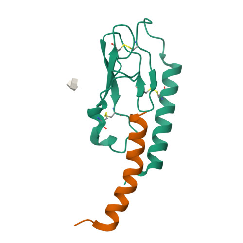
    3IOL

  • PubMed Abstract: 

    GLP-1 (glucagon-like peptide-1) is an incretin released from intestinal L-cells in response to food intake. Activation of the GLP-1 receptor potentiates the synthesis and release of insulin from pancreatic beta-cells in a glucose-dependent manner. The GLP-1 receptor belongs to class B of the G-protein-coupled receptors, a subfamily characterized by a large N-terminal extracellular ligand binding domain. Exendin-4 and GLP-1 are 50% identical, and exendin-4 is a full agonist with similar affinity and potency for the GLP-1 receptor. We recently solved the crystal structure of the GLP-1 receptor extracellular domain in complex with the competitive antagonist exendin-4(9-39). Interestingly, the isolated extracellular domain binds exendin-4 with much higher affinity than the endogenous agonist GLP-1. Here, we have solved the crystal structure of the extracellular domain in complex with GLP-1 to 2.1 Aresolution. The structure shows that important hydrophobic ligand-receptor interactions are conserved in agonist- and antagonist-bound forms of the extracellular domain, but certain residues in the ligand-binding site adopt a GLP-1-specific conformation. GLP-1 is a kinked but continuous alpha-helix from Thr(13) to Val(33) when bound to the extracellular domain. We supplemented the crystal structure with site-directed mutagenesis to link the structural information of the isolated extracellular domain with the binding properties of the full-length receptor. The data support the existence of differences in the binding modes of GLP-1 and exendin-4 on the full-length GLP-1 receptor.


  • Organizational Affiliation

    Department of GLP-1 and Obesity Biology, Novo Nordisk, 2760 Måløv, Denmark.


Macromolecules
Find similar proteins by:  (by identity cutoff)  |  3D Structure
Entity ID: 1
MoleculeChains Sequence LengthOrganismDetailsImage
Glucagon-like peptide 1 receptor126Homo sapiensMutation(s): 0 
Membrane Entity: Yes 
UniProt & NIH Common Fund Data Resources
Find proteins for P43220 (Homo sapiens)
Explore P43220 
Go to UniProtKB:  P43220
PHAROS:  P43220
GTEx:  ENSG00000112164 
Entity Groups  
Sequence Clusters30% Identity50% Identity70% Identity90% Identity95% Identity100% Identity
UniProt GroupP43220
Sequence Annotations
Expand
  • Reference Sequence
Find similar proteins by:  (by identity cutoff)  |  3D Structure
Entity ID: 2
MoleculeChains Sequence LengthOrganismDetailsImage
Glucagon31Homo sapiensMutation(s): 0 
Membrane Entity: Yes 
UniProt & NIH Common Fund Data Resources
Find proteins for P01275 (Homo sapiens)
Explore P01275 
Go to UniProtKB:  P01275
PHAROS:  P01275
GTEx:  ENSG00000115263 
Entity Groups  
Sequence Clusters30% Identity50% Identity70% Identity90% Identity95% Identity100% Identity
UniProt GroupP01275
Sequence Annotations
Expand
  • Reference Sequence
Small Molecules
Ligands 1 Unique
IDChains Name / Formula / InChI Key2D Diagram3D Interactions
10M
Query on 10M

Download Ideal Coordinates CCD File 
C [auth A]decyl 4-O-alpha-D-glucopyranosyl-1-thio-beta-D-glucopyranoside
C22 H42 O10 S
YZNNXXWNKQOETJ-HYLFJBCQSA-N
Experimental Data & Validation

Experimental Data

  • Method: X-RAY DIFFRACTION
  • Resolution: 2.10 Å
  • R-Value Free:  0.226 (Depositor), 0.240 (DCC) 
  • R-Value Work:  0.178 (Depositor), 0.190 (DCC) 
  • R-Value Observed: 0.181 (Depositor) 
Space Group: P 21 2 21
Unit Cell:
Length ( Å )Angle ( ˚ )
a = 35.67α = 90
b = 42.67β = 90
c = 95.09γ = 90
Software Package:
Software NamePurpose
PHASERphasing
REFMACrefinement
XDSdata reduction
XSCALEdata scaling

Structure Validation

View Full Validation Report


Ligand Structure Quality Assessment 

Created with Raphaël 2.3.0Worse 01 BetterLigand structure goodness of fit to experimental dataBest fitted 10MClick on this verticalbar to view details

Entry History 

Deposition Data

Revision History  (Full details and data files)

  • Version 1.0: 2009-10-27
    Type: Initial release
  • Version 1.1: 2011-07-13
    Changes: Advisory, Version format compliance
  • Version 1.2: 2018-05-23
    Changes: Advisory, Data collection
  • Version 1.3: 2023-11-01
    Changes: Advisory, Data collection, Database references, Derived calculations, Refinement description
  • Version 1.4: 2024-10-09
    Changes: Structure summary