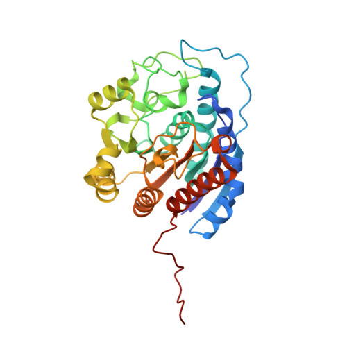
3SKK

Crystal structure of human arginase I in complex with the inhibitor FABH, Resolution 1.70 A, twinned structure


Experimental Data Snapshot

  • Method: X-RAY DIFFRACTION
  • Resolution: 1.70 Å
  • R-Value Free: 0.178 
  • R-Value Work: 0.136 
  • R-Value Observed: 0.140 

Starting Model: experimental
View more details

wwPDB Validation   3D Report Full Report


Ligand Structure Quality Assessment 


This is version 1.2 of the entry. See complete history


Literature

Binding of alpha , alpha-disubstituted amino acids to arginase suggests new avenues for inhibitor design.

Ilies, M.Di Costanzo, L.Dowling, D.P.Thorn, K.J.Christianson, D.W.

(2011) J Med Chem 54: 5432-5443

  • DOI: https://doi.org/10.1021/jm200443b
  • Primary Citation of Related Structures:  
    3GMZ, 3GN0, 3SJT, 3SKK, 3SL0, 3SL1

  • PubMed Abstract: 

    Arginase is a binuclear manganese metalloenzyme that hydrolyzes L-arginine to form L-ornithine and urea, and aberrant arginase activity is implicated in various diseases such as erectile dysfunction, asthma, atherosclerosis, and cerebral malaria. Accordingly, arginase inhibitors may be therapeutically useful. Continuing our efforts to expand the chemical space of arginase inhibitor design and inspired by the binding of 2-(difluoromethyl)-L-ornithine to human arginase I, we now report the first study of the binding of α,α-disubstituted amino acids to arginase. Specifically, we report the design, synthesis, and assay of racemic 2-amino-6-borono-2-methylhexanoic acid and racemic 2-amino-6-borono-2-(difluoromethyl)hexanoic acid. X-ray crystal structures of human arginase I and Plasmodium falciparum arginase complexed with these inhibitors reveal the exclusive binding of the L-stereoisomer; the additional α-substituent of each inhibitor is readily accommodated and makes new intermolecular interactions in the outer active site of each enzyme. Therefore, this work highlights a new region of the protein surface that can be targeted for additional affinity interactions, as well as the first comparative structural insights on inhibitor discrimination between a human and a parasitic arginase.


  • Organizational Affiliation

    Department of Chemistry, Drexel University, Philadelphia, Pennsylvania 19104-2875, United States.


Macromolecules
Find similar proteins by:  (by identity cutoff)  |  3D Structure
Entity ID: 1
MoleculeChains Sequence LengthOrganismDetailsImage
Arginase-1
A, B
322Homo sapiensMutation(s): 0 
Gene Names: ARG1
EC: 3.5.3.1
UniProt & NIH Common Fund Data Resources
Find proteins for P05089 (Homo sapiens)
Explore P05089 
Go to UniProtKB:  P05089
PHAROS:  P05089
GTEx:  ENSG00000118520 
Entity Groups  
Sequence Clusters30% Identity50% Identity70% Identity90% Identity95% Identity100% Identity
UniProt GroupP05089
Sequence Annotations
Expand
  • Reference Sequence
Experimental Data & Validation

Experimental Data

  • Method: X-RAY DIFFRACTION
  • Resolution: 1.70 Å
  • R-Value Free: 0.178 
  • R-Value Work: 0.136 
  • R-Value Observed: 0.140 
  • Space Group: P 3
Unit Cell:
Length ( Å )Angle ( ˚ )
a = 90.094α = 90
b = 90.094β = 90
c = 69.362γ = 120
Software Package:
Software NamePurpose
PHASERphasing
PHENIXrefinement
HKL-2000data reduction
SCALEPACKdata scaling

Structure Validation

View Full Validation Report


Ligand Structure Quality Assessment 


Entry History 

Deposition Data

Revision History  (Full details and data files)

  • Version 1.0: 2011-07-20
    Type: Initial release
  • Version 1.1: 2023-07-26
    Changes: Database references, Derived calculations
  • Version 1.2: 2023-09-13
    Changes: Data collection, Refinement description