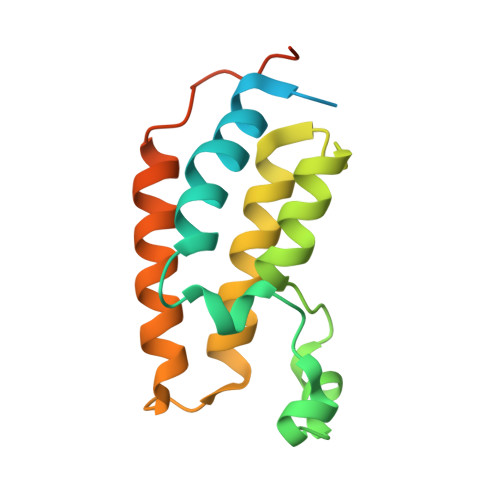
2YEK

Crystal Structure of the First Bromodomain of Human Brd2 with the inhibitor GSK525762 (IBET)


Experimental Data Snapshot

  • Method: X-RAY DIFFRACTION
  • Resolution: 1.98 Å
  • R-Value Free: 0.201 
  • R-Value Work: 0.155 
  • R-Value Observed: 0.158 

wwPDB Validation   3D Report Full Report


Ligand Structure Quality Assessment 


This is version 1.2 of the entry. See complete history


Literature

Discovery and Characterization of Small Molecule Inhibitors of the Bet Family Bromodomains.

Chung, C.W.Coste, H.White, J.H.Mirguet, O.Wilde, J.Gosmini, R.L.Delves, C.Magny, S.M.Woodward, R.Hughes, S.A.Boursier, E.V.Flynn, H.Bouillot, A.M.Bamborough, P.Brusq, J.M.Gellibert, F.J.Jones, E.J.Riou, A.M.Homes, P.Martin, S.L.Uings, I.J.Toum, J.Clement, C.A.Boullay, A.B.Grimley, R.L.Blandel, F.M.Prinjha, R.K.Lee, K.Kirilovsky, J.Nicodeme, E.

(2011) J Med Chem 54: 3827

  • DOI: https://doi.org/10.1021/jm200108t
  • Primary Citation of Related Structures:  
    2YDW, 2YEK, 2YEL, 2YEM

  • PubMed Abstract: 

    Epigenetic mechanisms of gene regulation have a profound role in normal development and disease processes. An integral part of this mechanism occurs through lysine acetylation of histone tails which are recognized by bromodomains. While the biological and structural characterization of many bromodomain containing proteins has advanced considerably, the therapeutic tractability of this protein family is only now becoming understood. This paper describes the discovery and molecular characterization of potent (nM) small molecule inhibitors that disrupt the function of the BET family of bromodomains (Brd2, Brd3, and Brd4). By using a combination of phenotypic screening, chemoproteomics, and biophysical studies, we have discovered that the protein-protein interactions between bromodomains and acetylated histones can be antagonized by selective small molecules that bind at the acetylated lysine recognition pocket. X-ray crystal structures of compounds bound into bromodomains of Brd2 and Brd4 elucidate the molecular interactions of binding and explain the precisely defined stereochemistry required for activity.


  • Organizational Affiliation

    Molecular Discovery Research, GlaxoSmithKline R&D, Stevenage SG5 4HT, UK. [email protected]


Macromolecules
Find similar proteins by:  (by identity cutoff)  |  3D Structure
Entity ID: 1
MoleculeChains Sequence LengthOrganismDetailsImage
BROMODOMAIN-CONTAINING PROTEIN 2
A, B, C
153Homo sapiensMutation(s): 0 
UniProt & NIH Common Fund Data Resources
Find proteins for P25440 (Homo sapiens)
Explore P25440 
Go to UniProtKB:  P25440
PHAROS:  P25440
GTEx:  ENSG00000204256 
Entity Groups  
Sequence Clusters30% Identity50% Identity70% Identity90% Identity95% Identity100% Identity
UniProt GroupP25440
Sequence Annotations
Expand
  • Reference Sequence
Small Molecules
Binding Affinity Annotations 
IDSourceBinding Affinity
EAM BindingDB:  2YEK Ki: min: 49, max: 150 (nM) from 4 assay(s)
Kd: min: 25, max: 230 (nM) from 8 assay(s)
IC50: min: 32.5, max: 810 (nM) from 4 assay(s)
PDBBind:  2YEK Kd: 61.3 (nM) from 1 assay(s)
Experimental Data & Validation

Experimental Data

  • Method: X-RAY DIFFRACTION
  • Resolution: 1.98 Å
  • R-Value Free: 0.201 
  • R-Value Work: 0.155 
  • R-Value Observed: 0.158 
  • Space Group: C 1 2 1
Unit Cell:
Length ( Å )Angle ( ˚ )
a = 116.243α = 90
b = 55.639β = 93.63
c = 66.231γ = 90
Software Package:
Software NamePurpose
REFMACrefinement
MOSFLMdata reduction
SCALAdata scaling
REFMACphasing

Structure Validation

View Full Validation Report


Ligand Structure Quality Assessment 


Entry History 

Deposition Data

  • Released Date: 2011-06-15 
  • Deposition Author(s): Chung, C.W.

Revision History  (Full details and data files)

  • Version 1.0: 2011-06-15
    Type: Initial release
  • Version 1.1: 2012-11-21
    Changes: Database references, Version format compliance
  • Version 1.2: 2024-05-08
    Changes: Data collection, Database references, Derived calculations, Other