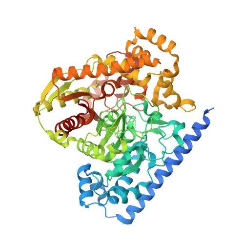
2VYA

Crystal Structure of fatty acid amide hydrolase conjugated with the drug-like inhibitor PF-750


Experimental Data Snapshot

  • Method: X-RAY DIFFRACTION
  • Resolution: 2.75 Å
  • R-Value Free: 0.239 
  • R-Value Work: 0.188 
  • R-Value Observed: 0.191 

Starting Model: experimental
View more details

wwPDB Validation   3D Report Full Report


Ligand Structure Quality Assessment 


This is version 1.3 of the entry. See complete history


Literature

Structure-Guided Inhibitor Design for Human Faah by Interspecies Active Site Conversion.

Mileni, M.Johnson, D.S.Wang, Z.Everdeen, D.S.Liimatta, M.Pabst, B.Bhattacharya, K.Nugent, R.A.Kamtekar, S.Cravatt, B.F.Ahn, K.Stevens, R.C.

(2008) Proc Natl Acad Sci U S A 105: 12820

  • DOI: https://doi.org/10.1073/pnas.0806121105
  • Primary Citation of Related Structures:  
    2VYA

  • PubMed Abstract: 

    The integral membrane enzyme fatty acid amide hydrolase (FAAH) hydrolyzes the endocannabinoid anandamide and related amidated signaling lipids. Genetic or pharmacological inactivation of FAAH produces analgesic, anxiolytic, and antiinflammatory phenotypes but not the undesirable side effects of direct cannabinoid receptor agonists, indicating that FAAH may be a promising therapeutic target. Structure-based inhibitor design has, however, been hampered by difficulties in expressing the human FAAH enzyme. Here, we address this problem by interconverting the active sites of rat and human FAAH using site-directed mutagenesis. The resulting humanized rat (h/r) FAAH protein exhibits the inhibitor sensitivity profiles of human FAAH but maintains the high-expression yield of the rat enzyme. We report a 2.75-A crystal structure of h/rFAAH complexed with an inhibitor, N-phenyl-4-(quinolin-3-ylmethyl)piperidine-1-carboxamide (PF-750), that shows strong preference for human FAAH. This structure offers compelling insights to explain the species selectivity of FAAH inhibitors, which should guide future drug design programs.


  • Organizational Affiliation

    The Skaggs Institute for Chemical Biology, La Jolla, CA 92037, USA.


Macromolecules
Find similar proteins by:  (by identity cutoff)  |  3D Structure
Entity ID: 1
MoleculeChains Sequence LengthOrganismDetailsImage
FATTY-ACID AMIDE HYDROLASE 1
A, B
587Rattus norvegicusMutation(s): 6 
EC: 3.5.1.4 (PDB Primary Data), 3.1.1 (UniProt), 3.5.1.99 (UniProt)
UniProt
Find proteins for P97612 (Rattus norvegicus)
Explore P97612 
Go to UniProtKB:  P97612
Entity Groups  
Sequence Clusters30% Identity50% Identity70% Identity90% Identity95% Identity100% Identity
UniProt GroupP97612
Sequence Annotations
Expand
  • Reference Sequence
Small Molecules
Ligands 3 Unique
IDChains Name / Formula / InChI Key2D Diagram3D Interactions
PF7
Query on PF7

Download Ideal Coordinates CCD File 
E [auth A],
I [auth B]
4-(quinolin-3-ylmethyl)piperidine-1-carboxylic acid
C16 H18 N2 O2
QUAGUFNCKDDJFZ-UHFFFAOYSA-N
CL
Query on CL

Download Ideal Coordinates CCD File 
H [auth B]CHLORIDE ION
Cl
VEXZGXHMUGYJMC-UHFFFAOYSA-M
UNX
Query on UNX

Download Ideal Coordinates CCD File 
C [auth A],
D [auth A],
F [auth B],
G [auth B]
UNKNOWN ATOM OR ION
X
Experimental Data & Validation

Experimental Data

  • Method: X-RAY DIFFRACTION
  • Resolution: 2.75 Å
  • R-Value Free: 0.239 
  • R-Value Work: 0.188 
  • R-Value Observed: 0.191 
  • Space Group: P 32 2 1
Unit Cell:
Length ( Å )Angle ( ˚ )
a = 103.69α = 90
b = 103.69β = 90
c = 253.87γ = 120
Software Package:
Software NamePurpose
REFMACrefinement
XDSdata reduction
XSCALEdata scaling
PHASERphasing

Structure Validation

View Full Validation Report


Ligand Structure Quality Assessment 


Entry History 

Revision History  (Full details and data files)

  • Version 1.0: 2008-09-09
    Type: Initial release
  • Version 1.1: 2011-07-13
    Changes: Advisory, Version format compliance
  • Version 1.2: 2023-12-13
    Changes: Data collection, Database references, Derived calculations, Other, Refinement description
  • Version 1.3: 2024-11-13
    Changes: Structure summary