2SAR

DETERMINATION AND RESTRAINED LEAST-SQUARES REFINEMENT OF THE CRYSTAL STRUCTURES OF RIBONUCLEASE SA AND ITS COMPLEX WITH 3'-GUANYLIC ACID AT 1.8 ANGSTROMS RESOLUTION


Experimental Data Snapshot

  • Method: X-RAY DIFFRACTION
  • Resolution: 1.80 Å
  • R-Value Observed: 0.175 

wwPDB Validation   3D Report Full Report


Ligand Structure Quality Assessment 

Created with Raphaël 2.3.0Worse 01 BetterLigand structure goodness of fit to experimental dataBest fitted 3GPClick on this verticalbar to view details

This is version 1.4 of the entry. See complete history


Literature

Determination and restrained least-squares refinement of the structures of ribonuclease Sa and its complex with 3'-guanylic acid at 1.8 A resolution.

Sevcik, J.Dodson, E.J.Dodson, G.G.

(1991) Acta Crystallogr B 47: 240-253

  • Primary Citation of Related Structures:  
    1SAR, 2SAR

  • PubMed Abstract: 

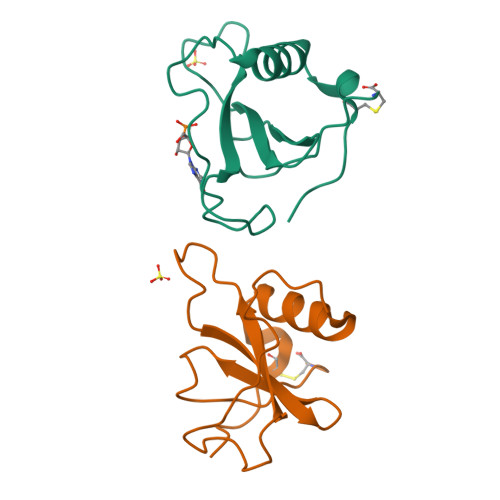
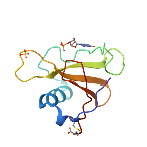
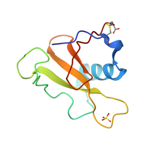
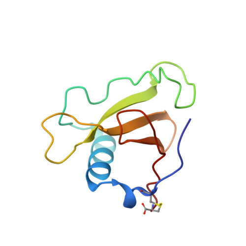
    The crystal structures of ribonuclease from Streptomyces aureofaciens (RNase Sa) and its complex with 3'-guanylic acid (guanosine 3'-monophosphate, 3'-GMP) have been determined by the method of isomorphous replacement. The atomic parameters have been refined by restrained least-squares minimization using data in the resolution range 10.0-1.8 A. All protein atoms and more than 230 water atoms in the two crystal structures have been refined to crystallographic R factors of 0.172 and 0.175 respectively. The estimated r.m.s. error in the atomic positions ranges from 0.2 A for well-defined atoms to about 0.5 A for more poorly defined atoms. There are two enzyme molecules in the asymmetric unit, built independently, and referred to as molecules A and B. The value of the average B factor for protein atoms in both structures is about 19 A2 and for water molecules about 35 A2. Electron density for the substrate analogue 3'-GMP was found only at the active site of molecule A. The density was very clear and the positions of all 3'-GMP atoms were refined with precision comparable to that of the protein.


  • Organizational Affiliation

    Institute of Molecular Biology, Slovak Academy of Sciences, Bratislava, Czechoslovakia.


Macromolecules
Find similar proteins by:  (by identity cutoff)  |  3D Structure
Entity ID: 1
MoleculeChains Sequence LengthOrganismDetailsImage
RIBONUCLEASE SA
A, B
96Kitasatospora aureofaciensMutation(s): 0 
EC: 4.6.1.24
UniProt
Find proteins for P05798 (Kitasatospora aureofaciens)
Explore P05798 
Go to UniProtKB:  P05798
Entity Groups  
Sequence Clusters30% Identity50% Identity70% Identity90% Identity95% Identity100% Identity
UniProt GroupP05798
Sequence Annotations
Expand
  • Reference Sequence
Experimental Data & Validation

Experimental Data

  • Method: X-RAY DIFFRACTION
  • Resolution: 1.80 Å
  • R-Value Observed: 0.175 
  • Space Group: P 21 21 21
Unit Cell:
Length ( Å )Angle ( ˚ )
a = 64.9α = 90
b = 78.32β = 90
c = 38.79γ = 90
Software Package:
Software NamePurpose
PROLSQrefinement

Structure Validation

View Full Validation Report


Ligand Structure Quality Assessment 

Created with Raphaël 2.3.0Worse 01 BetterLigand structure goodness of fit to experimental dataBest fitted 3GPClick on this verticalbar to view details

Entry History 

Deposition Data

Revision History  (Full details and data files)

  • Version 1.0: 1992-04-15
    Type: Initial release
  • Version 1.1: 2008-03-03
    Changes: Version format compliance
  • Version 1.2: 2011-07-13
    Changes: Version format compliance
  • Version 1.3: 2017-11-29
    Changes: Derived calculations, Other
  • Version 1.4: 2024-10-09
    Changes: Data collection, Database references, Derived calculations, Structure summary