2HV8 | pdb_00002hv8

Crystal structure of GTP-bound Rab11 in complex with FIP3


Experimental Data Snapshot

  • Method: X-RAY DIFFRACTION
  • Resolution: 1.86 Å
  • R-Value Free: 
    0.237 (Depositor), 0.240 (DCC) 
  • R-Value Work: 
    0.203 (Depositor), 0.200 (DCC) 
  • R-Value Observed: 
    0.205 (Depositor) 

Starting Model: experimental
View more details

wwPDB Validation   3D Report Full Report


Ligand Structure Quality Assessment 

Created with Raphaël 2.3.0Worse 01 BetterLigand structure goodness of fit to experimental dataBest fitted GTPClick on this verticalbar to view details

This is version 1.4 of the entry. See complete history


Literature

Structural Basis for Rab11-mediated Recruitment of FIP3 to Recycling Endosomes.

Eathiraj, S.Mishra, A.Prekeris, R.Lambright, D.G.

(2006) J Mol Biology 364: 121-135

  • DOI: https://doi.org/10.1016/j.jmb.2006.08.064
  • Primary Citation of Related Structures:  
    2HV8

  • PubMed Abstract: 

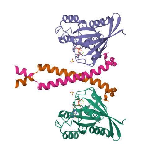
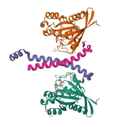
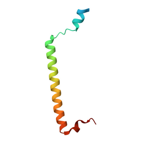
    The Rab11 GTPase regulates recycling of internalized plasma membrane receptors and is essential for completion of cytokinesis. A family of Rab11 interacting proteins (FIPs) that conserve a C-terminal Rab-binding domain (RBD) selectively recognize the active form of Rab11. Normal completion of cytokinesis requires a complex between Rab11 and FIP3. Here, we report the crystal structure and mutational analysis of a heterotetrameric complex between constitutively active Rab11 and a FIP3 construct that includes the RBD. Two Rab11 molecules bind to dyad symmetric sites at the C terminus of FIP3, which forms a non-canonical coiled-coiled dimer with a flared C terminus and hook region. The RBD overlaps with the coiled coil and extends through the C-terminal hook. Although FIP3 engages the switch and interswitch regions of Rab11, the mode of interaction differs significantly from that of other Rab-effector complexes. In particular, the switch II region undergoes a large structural rearrangement from an ordered but non-complementary active conformation to a remodeled conformation that facilitates the interaction with FIP3. Finally, we provide evidence that FIP3 can form homo-oligomers in cells, and that a critical determinant of Rab11 binding in vitro is necessary for FIP3 recruitment to recycling endosomes during cytokinesis.


  • Organizational Affiliation

    Program in Molecular Medicine and Department of Biochemistry & Molecular Pharmacology, University of Massachusetts Medical School, Worcester, MA 01605, USA.


Macromolecules
Find similar proteins by:  (by identity cutoff)  |  3D Structure
Entity ID: 1
MoleculeChains Sequence LengthOrganismDetailsImage
Ras-related protein Rab-11AA,
C [auth B],
E [auth C]
172Homo sapiensMutation(s): 1 
Gene Names: RAB11A
EC: 3.6.5.2
UniProt & NIH Common Fund Data Resources
Find proteins for P62491 (Homo sapiens)
Explore P62491 
Go to UniProtKB:  P62491
PHAROS:  P62491
GTEx:  ENSG00000103769 
Entity Groups  
Sequence Clusters30% Identity50% Identity70% Identity90% Identity95% Identity100% Identity
UniProt GroupP62491
Sequence Annotations
Expand
  • Reference Sequence
Find similar proteins by:  (by identity cutoff)  |  3D Structure
Entity ID: 2
MoleculeChains Sequence LengthOrganismDetailsImage
Rab11 family-interacting protein 3B [auth D],
D [auth E],
F
64Homo sapiensMutation(s): 0 
Gene Names: RAB11FIP3
UniProt & NIH Common Fund Data Resources
Find proteins for O75154 (Homo sapiens)
Explore O75154 
Go to UniProtKB:  O75154
PHAROS:  O75154
GTEx:  ENSG00000090565 
Entity Groups  
Sequence Clusters30% Identity50% Identity70% Identity90% Identity95% Identity100% Identity
UniProt GroupO75154
Sequence Annotations
Expand
  • Reference Sequence
Small Molecules
Ligands 5 Unique
IDChains Name / Formula / InChI Key2D Diagram3D Interactions
GTP
Query on GTP

Download Ideal Coordinates CCD File 
I [auth A],
N [auth B],
S [auth C]
GUANOSINE-5'-TRIPHOSPHATE
C10 H16 N5 O14 P3
XKMLYUALXHKNFT-UUOKFMHZSA-N
MES
Query on MES

Download Ideal Coordinates CCD File 
O [auth B]2-(N-MORPHOLINO)-ETHANESULFONIC ACID
C6 H13 N O4 S
SXGZJKUKBWWHRA-UHFFFAOYSA-N
SO4
Query on SO4

Download Ideal Coordinates CCD File 
H [auth A],
L [auth B],
M [auth B],
Q [auth C],
R [auth C]
SULFATE ION
O4 S
QAOWNCQODCNURD-UHFFFAOYSA-L
2ME
Query on 2ME

Download Ideal Coordinates CCD File 
J [auth B]METHOXYETHANE
C3 H8 O
XOBKSJJDNFUZPF-UHFFFAOYSA-N
MG
Query on MG

Download Ideal Coordinates CCD File 
G [auth A],
K [auth B],
P [auth C]
MAGNESIUM ION
Mg
JLVVSXFLKOJNIY-UHFFFAOYSA-N
Experimental Data & Validation

Experimental Data

  • Method: X-RAY DIFFRACTION
  • Resolution: 1.86 Å
  • R-Value Free:  0.237 (Depositor), 0.240 (DCC) 
  • R-Value Work:  0.203 (Depositor), 0.200 (DCC) 
  • R-Value Observed: 0.205 (Depositor) 
Space Group: C 1 2 1
Unit Cell:
Length ( Å )Angle ( ˚ )
a = 218.291α = 90
b = 52.601β = 105.56
c = 70.679γ = 90
Software Package:
Software NamePurpose
REFMACrefinement
DENZOdata reduction
SCALEPACKdata scaling
AMoREphasing

Structure Validation

View Full Validation Report


Ligand Structure Quality Assessment 

Created with Raphaël 2.3.0Worse 01 BetterLigand structure goodness of fit to experimental dataBest fitted GTPClick on this verticalbar to view details

Entry History 

Deposition Data

Revision History  (Full details and data files)

  • Version 1.0: 2006-11-21
    Type: Initial release
  • Version 1.1: 2008-05-01
    Changes: Version format compliance
  • Version 1.2: 2011-07-13
    Changes: Version format compliance
  • Version 1.3: 2021-10-20
    Changes: Database references, Derived calculations
  • Version 1.4: 2023-08-30
    Changes: Data collection, Refinement description