2BIX

Crystal structure of apocarotenoid cleavage oxygenase from Synechocystis, Fe-free apoenzyme


Experimental Data Snapshot

  • Method: X-RAY DIFFRACTION
  • Resolution: 2.68 Å
  • R-Value Free: 0.243 
  • R-Value Work: 0.210 
  • R-Value Observed: 0.210 

wwPDB Validation   3D Report Full Report


Ligand Structure Quality Assessment 

Created with Raphaël 2.3.0Worse 01 BetterLigand structure goodness of fit to experimental dataBest fitted C8EClick on this verticalbar to view details

This is version 1.3 of the entry. See complete history


Literature

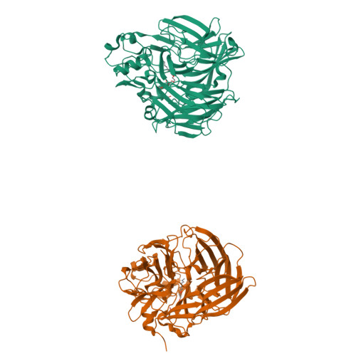
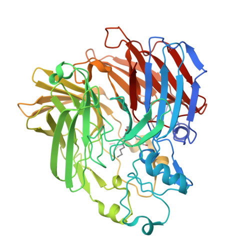
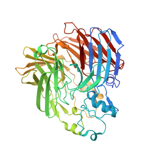
The Structure of a Retinal-Forming Carotenoid Oxygenase

Kloer, D.P.Ruch, S.Al-Babili, S.Beyer, P.Schulz, G.E.

(2005) Science 308: 267

  • DOI: https://doi.org/10.1126/science.1108965
  • Primary Citation of Related Structures:  
    2BIW, 2BIX

  • PubMed Abstract: 

    Enzymes that produce retinal and related apocarotenoids constitute a sequence- and thus structure-related family, a member of which was analyzed by x-ray diffraction. This member is an oxygenase and contains an Fe2+-4-His arrangement at the axis of a seven-bladed beta-propeller chain fold covered by a dome formed by six large loops. The Fe2+ is accessible through a long nonpolar tunnel that holds a carotenoid derivative in one of the crystals. On binding, three consecutive double bonds of this carotenoid changed from a straight all-trans to a cranked cis-trans-cis conformation. The remaining trans bond is located at the dioxygen-ligated Fe2+ and cleaved by oxygen.


  • Organizational Affiliation

    Institut für Organische Chemie und Biochemie, Albert-Ludwigs-Universität, Albertstrasse 21, 79104 Freiburg im Breisgau, Germany.


Macromolecules
Find similar proteins by:  (by identity cutoff)  |  3D Structure
Entity ID: 1
MoleculeChains Sequence LengthOrganismDetailsImage
APOCAROTENOID-CLEAVING OXYGENASE
A, B
490Synechocystis sp. PCC 6803Mutation(s): 0 
EC: 1.13.11.75
UniProt
Find proteins for P74334 (Synechocystis sp. (strain ATCC 27184 / PCC 6803 / Kazusa))
Explore P74334 
Go to UniProtKB:  P74334
Entity Groups  
Sequence Clusters30% Identity50% Identity70% Identity90% Identity95% Identity100% Identity
UniProt GroupP74334
Sequence Annotations
Expand
  • Reference Sequence
Experimental Data & Validation

Experimental Data

  • Method: X-RAY DIFFRACTION
  • Resolution: 2.68 Å
  • R-Value Free: 0.243 
  • R-Value Work: 0.210 
  • R-Value Observed: 0.210 
  • Space Group: P 41 21 2
Unit Cell:
Length ( Å )Angle ( ˚ )
a = 122.928α = 90
b = 122.928β = 90
c = 205.56γ = 90
Software Package:
Software NamePurpose
TNTrefinement
XDSdata reduction
XSCALEdata scaling
SHARPphasing

Structure Validation

View Full Validation Report


Ligand Structure Quality Assessment 

Created with Raphaël 2.3.0Worse 01 BetterLigand structure goodness of fit to experimental dataBest fitted C8EClick on this verticalbar to view details

Entry History 

Deposition Data

Revision History  (Full details and data files)

  • Version 1.0: 2005-04-14
    Type: Initial release
  • Version 1.1: 2011-05-08
    Changes: Version format compliance
  • Version 1.2: 2011-07-13
    Changes: Version format compliance
  • Version 1.3: 2024-05-08
    Changes: Data collection, Database references, Other