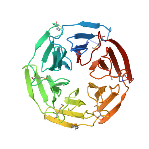
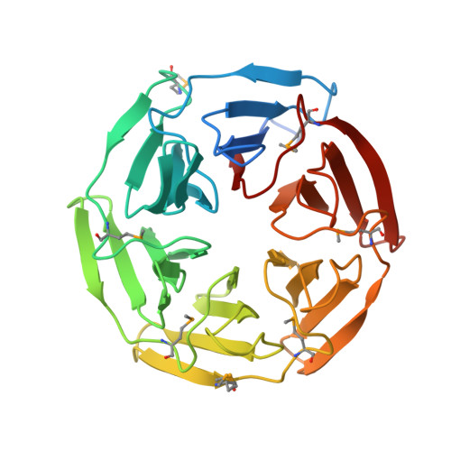
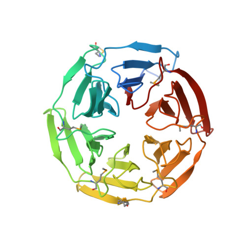
1ZGK

1.35 angstrom structure of the Kelch domain of Keap1


Experimental Data Snapshot

  • Method: X-RAY DIFFRACTION
  • Resolution: 1.35 Å
  • R-Value Free: 0.133 
  • R-Value Work: 0.111 
  • R-Value Observed: 0.112 

wwPDB Validation   3D Report Full Report


This is version 1.3 of the entry. See complete history


Literature

Conserved solvent and side-chain interactions in the 1.35 Angstrom structure of the Kelch domain of Keap1.

Beamer, L.J.Li, X.Bottoms, C.A.Hannink, M.

(2005) Acta Crystallogr D Biol Crystallogr 61: 1335-1342

  • DOI: https://doi.org/10.1107/S0907444905022626
  • Primary Citation of Related Structures:  
    1ZGK

  • PubMed Abstract: 

    The Kelch repeat is a common sequence motif in eukaryotic genomes and is approximately 50 amino acids in length. The structure of the Kelch domain of the human Keap1 protein has previously been determined at 1.85 Angstrom, showing that each Kelch repeat forms one blade of a six-bladed beta-propeller. Here, use of 1.35 Angstrom SAD data for de novo structure determination of the Kelch domain and for refinement at atomic resolution is described. The high quality and resolution of the diffraction data and phase information allows a detailed analysis of the role of solvent in the structure of the Kelch repeat. Ten structurally conserved water molecules are identified in each blade of the Kelch beta-propeller. These appear to play distinct structural roles that include lining the central channel of the propeller, interacting with residues in loops between strands of the blade and making contacts with conserved residues in the Kelch repeat. Furthermore, we identify a conserved C-H...pi hydrogen bond between two key residues in the consensus Kelch repeat. This analysis extends our understanding of the structural roles of conserved residues in the Kelch repeat and highlights the potential role of solvent in maintaining the fold of this common eukaryotic structural motif.


  • Organizational Affiliation

    Department of Biochemistry, University of Missouri-Columbia, Columbia, MO 65211, USA. [email protected]


Macromolecules
Find similar proteins by:  (by identity cutoff)  |  3D Structure
Entity ID: 1
MoleculeChains Sequence LengthOrganismDetailsImage
Kelch-like ECH-associated protein 1308Homo sapiensMutation(s): 7 
Gene Names: KEAP1KIAA0132
UniProt & NIH Common Fund Data Resources
Find proteins for Q14145 (Homo sapiens)
Explore Q14145 
Go to UniProtKB:  Q14145
PHAROS:  Q14145
GTEx:  ENSG00000079999 
Entity Groups  
Sequence Clusters30% Identity50% Identity70% Identity90% Identity95% Identity100% Identity
UniProt GroupQ14145
Sequence Annotations
Expand
  • Reference Sequence
Small Molecules
Modified Residues  1 Unique
IDChains TypeFormula2D DiagramParent
MSE
Query on MSE
A
L-PEPTIDE LINKINGC5 H11 N O2 SeMET
Experimental Data & Validation

Experimental Data

  • Method: X-RAY DIFFRACTION
  • Resolution: 1.35 Å
  • R-Value Free: 0.133 
  • R-Value Work: 0.111 
  • R-Value Observed: 0.112 
  • Space Group: P 65 2 2
Unit Cell:
Length ( Å )Angle ( ˚ )
a = 85.745α = 90
b = 85.745β = 90
c = 147.818γ = 120
Software Package:
Software NamePurpose
REFMACrefinement
HKL-2000data reduction
CCP4data scaling
SOLVEphasing

Structure Validation

View Full Validation Report


Entry History 

Deposition Data

Revision History  (Full details and data files)

  • Version 1.0: 2005-10-04
    Type: Initial release
  • Version 1.1: 2008-04-30
    Changes: Version format compliance
  • Version 1.2: 2011-07-13
    Changes: Version format compliance
  • Version 1.3: 2024-11-20
    Changes: Data collection, Database references, Derived calculations, Structure summary