1VDC

STRUCTURE OF NADPH DEPENDENT THIOREDOXIN REDUCTASE


Experimental Data Snapshot

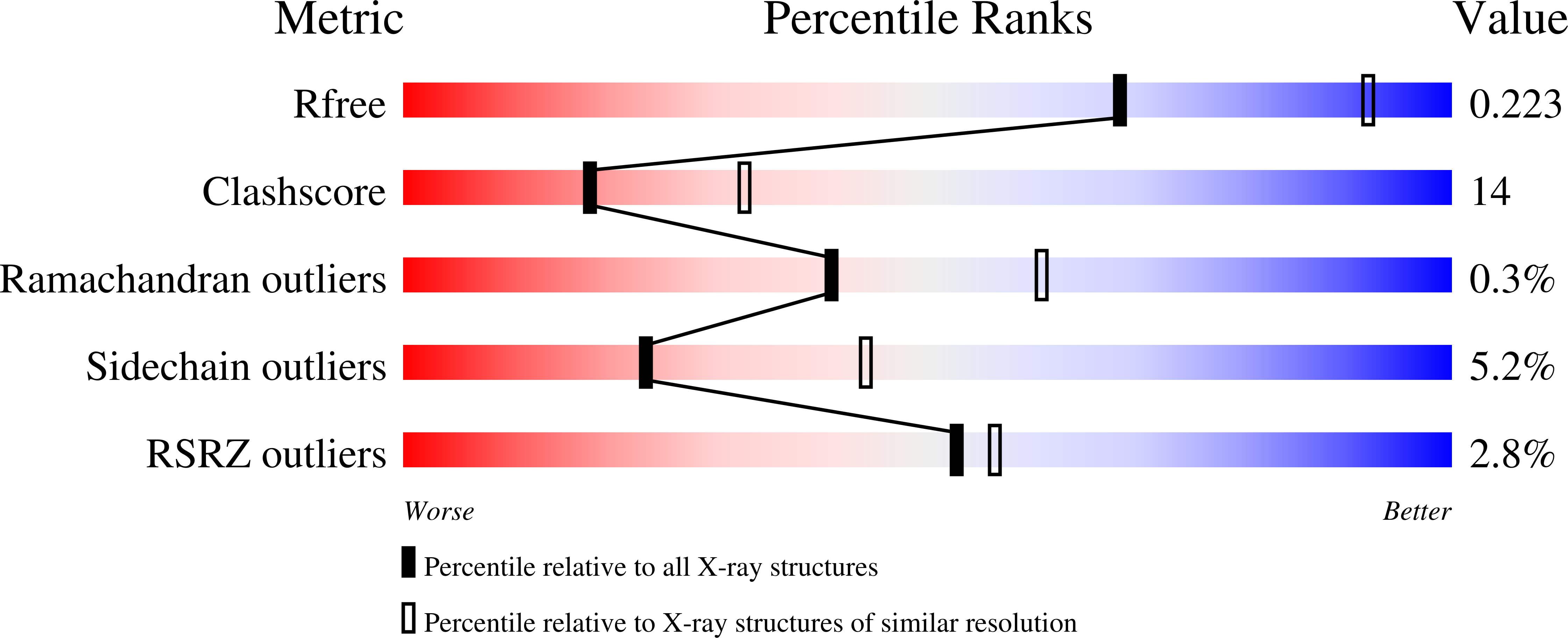
  • Method: X-RAY DIFFRACTION
  • Resolution: 2.50 Å
  • R-Value Free: 0.243 
  • R-Value Work: 0.188 
  • R-Value Observed: 0.188 

Starting Model: experimental
View more details

wwPDB Validation   3D Report Full Report


Ligand Structure Quality Assessment 


This is version 1.4 of the entry. See complete history


Literature

Crystal structure of Arabidopsis thaliana NADPH dependent thioredoxin reductase at 2.5 A resolution.

Dai, S.Saarinen, M.Ramaswamy, S.Meyer, Y.Jacquot, J.P.Eklund, H.

(1996) J Mol Biol 264: 1044-1057

  • DOI: https://doi.org/10.1006/jmbi.1996.0695
  • Primary Citation of Related Structures:  
    1VDC

  • PubMed Abstract: 

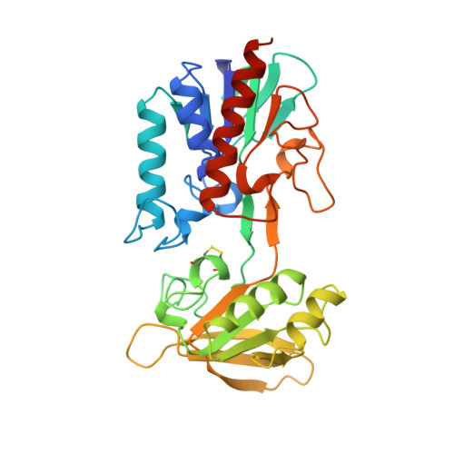
    Thioredoxin exists in all organisms and is responsible for the hydrogen transfer to important enzymes for ribonucleotide reduction and the reduction of methionine sulphoxide and sulphate. Thioredoxins have also been shown to regulate enzyme activity in plants and are also involved in the regulation of transcription factors and several other regulatory activities. Thioredoxin is reduced by the flavoenzyme thioredoxin reductase using NADPH. We have now determined the first structure of a eukaryotic thioredoxin reductase, from the plant Arabidopsis thaliana, at 2.5 A resolution. The dimeric A. thaliana thioredoxin reductase is structurally similar to that of the Escherichia coli enzyme, and most differences occur in the loops. Because the plant and E. coli enzymes have the same architecture, with the same dimeric structure and the same position of the redox active disulphide bond, a similar mechanism that involves very large domain rotations is likely for the two enzymes. The subunit is divided into two domains, one that binds FAD and one that binds NADPH. The relative positions of the domains in A. thaliana thioredoxin reductase differ from those of the E. coli reductase. When the FAD domains are superimposed, the NADPH domain of A. thaliana thioredoxin reductase must be rotated by 8 degrees to superimpose on the corresponding domain of the E. coli enzyme. The domain rotation we now observe is much smaller than necessary for the thioredoxin reduction cycle.


  • Organizational Affiliation

    Department of Molecular Biology, Swedish University of Agricultural Sciences, Uppsala, Sweden.


Macromolecules
Find similar proteins by:  (by identity cutoff)  |  3D Structure
Entity ID: 1
MoleculeChains Sequence LengthOrganismDetailsImage
NADPH DEPENDENT THIOREDOXIN REDUCTASE333Arabidopsis thalianaMutation(s): 0 
Gene Names: ATTHIREDB
EC: 1.6.4.5 (PDB Primary Data), 1.8.1.9 (UniProt)
UniProt
Find proteins for Q39243 (Arabidopsis thaliana)
Explore Q39243 
Go to UniProtKB:  Q39243
Entity Groups  
Sequence Clusters30% Identity50% Identity70% Identity90% Identity95% Identity100% Identity
UniProt GroupQ39243
Sequence Annotations
Expand
  • Reference Sequence
Experimental Data & Validation

Experimental Data

  • Method: X-RAY DIFFRACTION
  • Resolution: 2.50 Å
  • R-Value Free: 0.243 
  • R-Value Work: 0.188 
  • R-Value Observed: 0.188 
  • Space Group: P 41 21 2
Unit Cell:
Length ( Å )Angle ( ˚ )
a = 89.974α = 90
b = 89.974β = 90
c = 175.626γ = 90
Software Package:
Software NamePurpose
DENZOdata reduction
SCALEPACKdata scaling
X-PLORmodel building
X-PLORrefinement
X-PLORphasing

Structure Validation

View Full Validation Report


Ligand Structure Quality Assessment 


Entry History 

Deposition Data

Revision History  (Full details and data files)

  • Version 1.0: 1997-03-12
    Type: Initial release
  • Version 1.1: 2008-03-24
    Changes: Version format compliance
  • Version 1.2: 2011-07-13
    Changes: Derived calculations, Version format compliance
  • Version 1.3: 2023-08-09
    Changes: Data collection, Database references, Derived calculations, Refinement description
  • Version 1.4: 2024-10-16
    Changes: Data collection, Structure summary