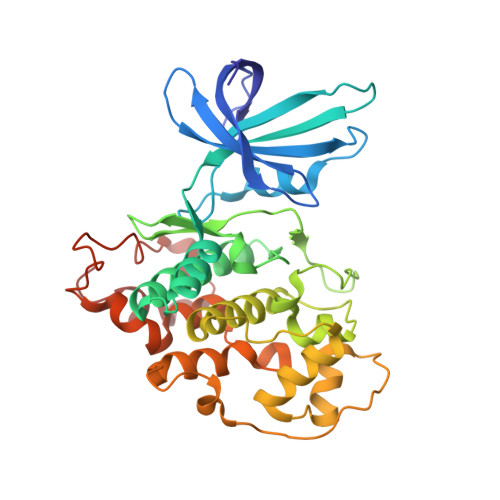
1UV5

GLYCOGEN SYNTHASE KINASE 3 BETA COMPLEXED WITH 6-BROMOINDIRUBIN-3'-OXIME


Experimental Data Snapshot

  • Method: X-RAY DIFFRACTION
  • Resolution: 2.80 Å
  • R-Value Free: 0.226 
  • R-Value Work: 0.193 
  • R-Value Observed: 0.193 

Starting Model: experimental
View more details

wwPDB Validation   3D Report Full Report


Ligand Structure Quality Assessment 


This is version 1.1 of the entry. See complete history


Literature

Gsk-3-Selective Inhibitors Derived from Tyrian Purple Indurubins

Meijer, L.Skaltsounis, A.-L.Magiatis, P.Polychronopoulous, P.Knockaert, M.Leost, M.Ryan, X.P.Vonica, C.A.Brivanlou, A.Dajani, R.Crovace, C.Tarricone, C.Musacchio, A.Roe, S.M.Pearl, L.H.Greengard, P.

(2003) Chem Biol 10: 1255

  • DOI: https://doi.org/10.1016/j.chembiol.2003.11.010
  • Primary Citation of Related Structures:  
    1UV5

  • PubMed Abstract: 

    Gastropod mollusks have been used for over 2500 years to produce the "Tyrian purple" dye made famous by the Phoenicians. This dye is constituted of mixed bromine-substituted indigo and indirubin isomers. Among these, the new natural product 6-bromoindirubin and its synthetic, cell-permeable derivative, 6-bromoindirubin-3'-oxime (BIO), display remarkable selective inhibition of glycogen synthase kinase-3 (GSK-3). Cocrystal structure of GSK-3beta/BIO and CDK5/p25/indirubin-3'-oxime were resolved, providing a detailed view of indirubins' interactions within the ATP binding pocket of these kinases. BIO but not 1-methyl-BIO, its kinase inactive analog, also inhibited the phosphorylation on Tyr276/216, a GSK-3alpha/beta activation site. BIO but not 1-methyl-BIO reduced beta-catenin phosphorylation on a GSK-3-specific site in cellular models. BIO but not 1-methyl-BIO closely mimicked Wnt signaling in Xenopus embryos. 6-bromoindirubins thus provide a new scaffold for the development of selective and potent pharmacological inhibitors of GSK-3.


  • Organizational Affiliation

    CNRS, Cell Cycle Group, Station Biologique, BP 74, 29682 Roscoff cedex, Bretagne, France. [email protected]


Macromolecules
Find similar proteins by:  (by identity cutoff)  |  3D Structure
Entity ID: 1
MoleculeChains Sequence LengthOrganismDetailsImage
GLYCOGEN SYNTHASE KINASE-3 BETA350Homo sapiensMutation(s): 0 
EC: 2.7.1.37 (PDB Primary Data), 2.7.11.1 (UniProt), 2.7.11.26 (UniProt)
UniProt & NIH Common Fund Data Resources
Find proteins for P49841 (Homo sapiens)
Explore P49841 
Go to UniProtKB:  P49841
PHAROS:  P49841
GTEx:  ENSG00000082701 
Entity Groups  
Sequence Clusters30% Identity50% Identity70% Identity90% Identity95% Identity100% Identity
UniProt GroupP49841
Sequence Annotations
Expand
  • Reference Sequence
Small Molecules
Binding Affinity Annotations 
IDSourceBinding Affinity
CL BindingDB:  1UV5 IC50: 6530 (nM) from 1 assay(s)
EC50: 3.00e+6 (nM) from 1 assay(s)
BRW BindingDB:  1UV5 IC50: min: 5, max: 1500 (nM) from 2 assay(s)
PDBBind:  1UV5 IC50: 5 (nM) from 1 assay(s)
Experimental Data & Validation

Experimental Data

  • Method: X-RAY DIFFRACTION
  • Resolution: 2.80 Å
  • R-Value Free: 0.226 
  • R-Value Work: 0.193 
  • R-Value Observed: 0.193 
  • Space Group: P 43 21 2
Unit Cell:
Length ( Å )Angle ( ˚ )
a = 98.333α = 90
b = 98.333β = 90
c = 198.017γ = 90
Software Package:
Software NamePurpose
CNSrefinement
MOSFLMdata reduction
SCALAdata scaling
AMoREphasing

Structure Validation

View Full Validation Report


Ligand Structure Quality Assessment 


Entry History 

Deposition Data

Revision History  (Full details and data files)

  • Version 1.0: 2004-01-29
    Type: Initial release
  • Version 1.1: 2023-12-13
    Changes: Data collection, Database references, Derived calculations, Other, Refinement description