1LOW

X-ray structure of the H40A mutant of Ribonuclease T1 complexed with 3'-guanosine monophosphate


Experimental Data Snapshot

  • Method: X-RAY DIFFRACTION
  • Resolution: 1.90 Å
  • R-Value Free: 
    0.229 (Depositor) 
  • R-Value Work: 
    0.206 (Depositor), 0.200 (DCC) 
  • R-Value Observed: 
    0.210 (Depositor) 

Starting Model: experimental
View more details

wwPDB Validation   3D Report Full Report


Ligand Structure Quality Assessment 

Created with Raphaël 2.3.0Worse 01 BetterLigand structure goodness of fit to experimental dataBest fitted 3GPClick on this verticalbar to view details

This is version 1.5 of the entry. See complete history


Literature

A nucleophile activation dyad in ribonucleases. A combined X-ray crystallographic/ab initio quantum chemical study

Mignon, P.Steyaert, J.Loris, R.Geerlings, P.Loverix, S.

(2002) J Biol Chem 277: 36770-36774

  • DOI: https://doi.org/10.1074/jbc.M206461200
  • Primary Citation of Related Structures:  
    1LOV, 1LOW, 1LOY

  • PubMed Abstract: 

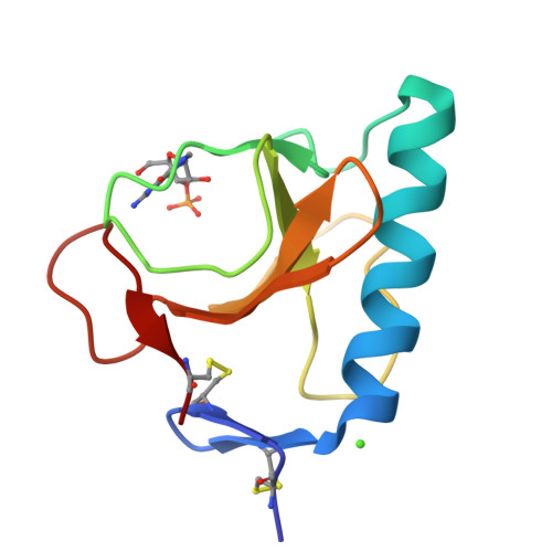
    Ribonucleases (RNases) catalyze the cleavage of the phosphodiester bond in RNA up to 10(15)-fold, as compared with the uncatalyzed reaction. High resolution crystal structures of these enzymes in complex with 3'-mononucleotide substrates demonstrate the accommodation of the nucleophilic 2'-OH group in a binding pocket comprising the catalytic base (glutamate or histidine) and a charged hydrogen bond donor (lysine or histidine). Ab initio quantum chemical calculations performed on such Michaelis complexes of the mammalian RNase A (EC ) and the microbial RNase T(1) (EC ) show negative charge build up on the 2'-oxygen upon substrate binding. The increased nucleophilicity results from stronger hydrogen bonding to the catalytic base, which is mediated by a hydrogen bond from the charged donor. This hitherto unrecognized catalytic dyad in ribonucleases constitutes a general mechanism for nucleophile activation in both enzymic and RNA-catalyzed phosphoryl transfer reactions.


  • Organizational Affiliation

    Eenheid Algemene Chemie (ALGC), Faculteit Wetenschappen, Vrije Universiteit Brussel, Pleinlaan 2, 1050 Brussels, Belgium.


Macromolecules
Find similar proteins by:  (by identity cutoff)  |  3D Structure
Entity ID: 1
MoleculeChains Sequence LengthOrganismDetailsImage
Guanyl-specific ribonuclease T1104Aspergillus oryzaeMutation(s): 1 
EC: 3.1.27.3 (PDB Primary Data), 4.6.1.24 (UniProt)
UniProt
Find proteins for P00651 (Aspergillus oryzae (strain ATCC 42149 / RIB 40))
Explore P00651 
Go to UniProtKB:  P00651
Entity Groups  
Sequence Clusters30% Identity50% Identity70% Identity90% Identity95% Identity100% Identity
UniProt GroupP00651
Sequence Annotations
Expand
  • Reference Sequence
Experimental Data & Validation

Experimental Data

  • Method: X-RAY DIFFRACTION
  • Resolution: 1.90 Å
  • R-Value Free:  0.229 (Depositor) 
  • R-Value Work:  0.206 (Depositor), 0.200 (DCC) 
  • R-Value Observed: 0.210 (Depositor) 
Space Group: P 21 21 21
Unit Cell:
Length ( Å )Angle ( ˚ )
a = 40.217α = 90
b = 45.685β = 90
c = 50.136γ = 90
Software Package:
Software NamePurpose
DENZOdata reduction
SCALEPACKdata scaling
CNSrefinement
CNSphasing

Structure Validation

View Full Validation Report


Ligand Structure Quality Assessment 

Created with Raphaël 2.3.0Worse 01 BetterLigand structure goodness of fit to experimental dataBest fitted 3GPClick on this verticalbar to view details

Entry History 

Deposition Data

Revision History  (Full details and data files)

  • Version 1.0: 2002-08-21
    Type: Initial release
  • Version 1.1: 2008-04-28
    Changes: Version format compliance
  • Version 1.2: 2011-07-13
    Changes: Version format compliance
  • Version 1.3: 2021-10-27
    Changes: Database references, Derived calculations
  • Version 1.4: 2023-08-16
    Changes: Data collection, Refinement description
  • Version 1.5: 2024-10-30
    Changes: Structure summary