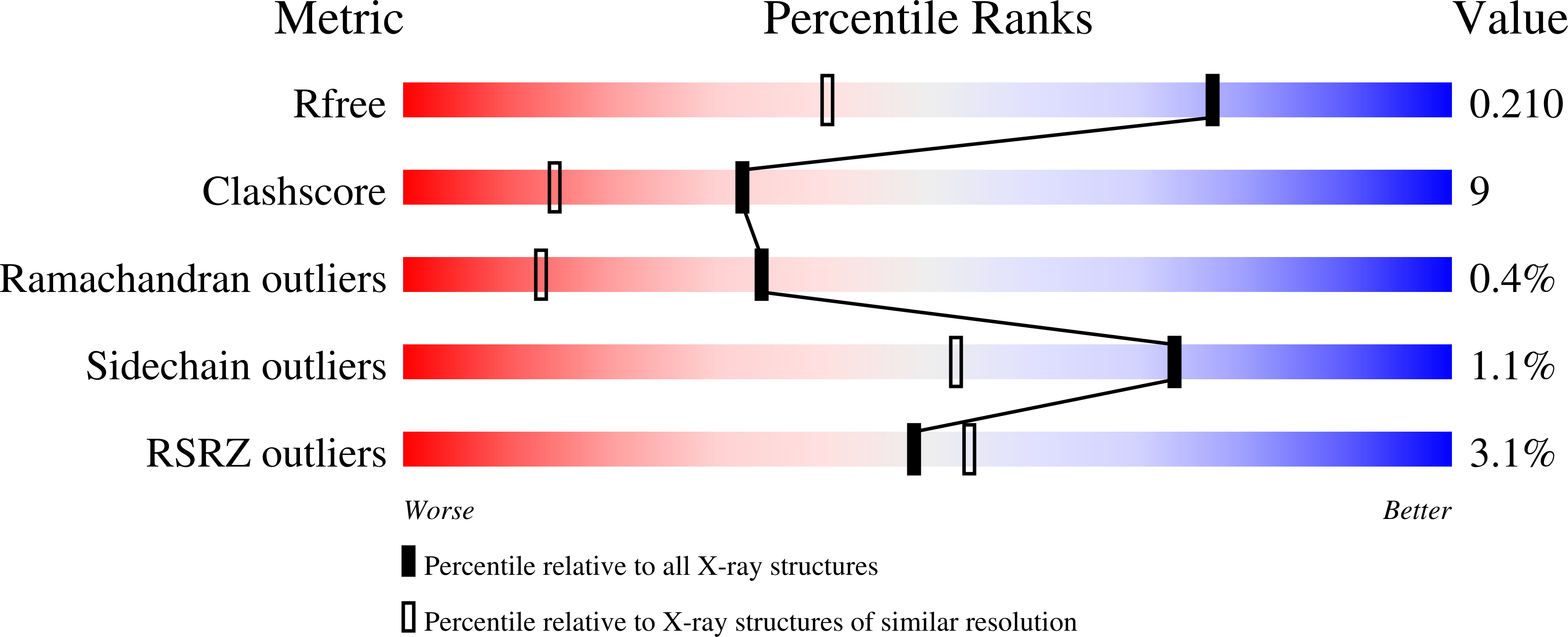
1HXK

GOLGI ALPHA-MANNOSIDASE II IN COMPLEX WITH DEOXYMANNOJIRIMICIN


Experimental Data Snapshot

  • Method: X-RAY DIFFRACTION
  • Resolution: 1.50 Å
  • R-Value Free: 0.216 
  • R-Value Work: 0.197 

Starting Model: experimental
View more details

wwPDB Validation   3D Report Full Report


Ligand Structure Quality Assessment 

Created with Raphaël 2.3.0Worse 01 BetterLigand structure goodness of fit to experimental dataBest fitted NAGClick on this verticalbar to view detailsBest fitted DMJClick on this verticalbar to view details

This is version 1.5 of the entry. See complete history


Literature

Structure of Golgi alpha-mannosidase II: a target for inhibition of growth and metastasis of cancer cells.

van den Elsen, J.M.Kuntz, D.A.Rose, D.R.

(2001) EMBO J 20: 3008-3017

  • DOI: https://doi.org/10.1093/emboj/20.12.3008
  • Primary Citation of Related Structures:  
    1HTY, 1HWW, 1HXK

  • PubMed Abstract: 

    Golgi alpha-mannosidase II, a key enzyme in N-glycan processing, is a target in the development of anti- cancer therapies. The crystal structure of Drosophila Golgi alpha-mannosidase II in the absence and presence of the anti-cancer agent swainsonine and the inhibitor deoxymannojirimycin reveals a novel protein fold with an active site zinc intricately involved both in the substrate specificity of the enzyme and directly in the catalytic mechanism. Identification of a putative GlcNAc binding pocket in the vicinity of the active site cavity provides a model for the binding of the GlcNAcMan(5)GlcNAc(2) substrate and the consecutive hydrolysis of the alpha1,6- and alpha1,3-linked mannose residues. The enzyme-inhibitor interactions observed provide insight into the catalytic mechanism, opening the door to the design of novel inhibitors of alpha-mannosidase II.


  • Organizational Affiliation

    Ontario Cancer Institute and Department of Medical Biophysics, University of Toronto, 610 University Avenue, Toronto, Ontario, Canada.


Macromolecules
Find similar proteins by:  (by identity cutoff)  |  3D Structure
Entity ID: 1
MoleculeChains Sequence LengthOrganismDetailsImage
ALPHA-MANNOSIDASE II1,015Drosophila melanogasterMutation(s): 0 
EC: 3.2.1.114
UniProt
Find proteins for Q24451 (Drosophila melanogaster)
Explore Q24451 
Go to UniProtKB:  Q24451
Entity Groups  
Sequence Clusters30% Identity50% Identity70% Identity90% Identity95% Identity100% Identity
UniProt GroupQ24451
Glycosylation
Glycosylation Sites: 1Go to GlyGen: Q24451-1
Sequence Annotations
Expand
  • Reference Sequence
Experimental Data & Validation

Experimental Data

  • Method: X-RAY DIFFRACTION
  • Resolution: 1.50 Å
  • R-Value Free: 0.216 
  • R-Value Work: 0.197 
  • Space Group: P 21 21 21
Unit Cell:
Length ( Å )Angle ( ˚ )
a = 68.814α = 90
b = 109.616β = 90
c = 138.281γ = 90
Software Package:
Software NamePurpose
CNSrefinement
DENZOdata reduction
SCALEPACKdata scaling
CNSphasing

Structure Validation

View Full Validation Report


Ligand Structure Quality Assessment 

Created with Raphaël 2.3.0Worse 01 BetterLigand structure goodness of fit to experimental dataBest fitted NAGClick on this verticalbar to view detailsBest fitted DMJClick on this verticalbar to view details

Entry History 

Deposition Data

Revision History  (Full details and data files)

  • Version 1.0: 2002-01-16
    Type: Initial release
  • Version 1.1: 2008-04-27
    Changes: Version format compliance
  • Version 1.2: 2011-07-13
    Changes: Non-polymer description, Version format compliance
  • Version 1.3: 2020-07-29
    Type: Remediation
    Reason: Carbohydrate remediation
    Changes: Data collection, Derived calculations, Structure summary
  • Version 1.4: 2023-08-09
    Changes: Data collection, Database references, Refinement description, Structure summary
  • Version 1.5: 2024-11-06
    Changes: Structure summary